Grace Davidson gave birth to a baby girl two years after her sister’s womb was transplanted into her body.
Category: biotech/medical – Page 142
First-of-Its-Kind Study Identifies How The Brain Could Thwart Alzheimer’s
Training the brain’s immune system to recognize and clear toxic material is rapidly emerging as an promising way to put the brakes on Alzheimer’s disease. Unfortunately researchers haven’t been clear on how this method of protection operates on a cellular level.
An international team of researchers analyzed brain samples taken from people who had died with Alzheimer’s, some of whom had also received approved Alzheimer’s immunotherapy treatments. The therapies encourage cleaning cells called microglia to attack the clumps of amyloid-beta proteins that are thought to be involved in neurodegeneration.
Microglia responses to amyloid beta can lead to inflammation, which in turn risks damage to brain tissues. The researchers wanted to know why immunotherapy turned microglia into ruthless cleaning machines in some cases but not others.
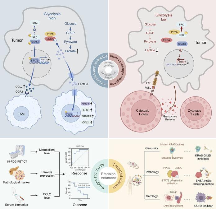
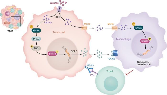
Abstract: Why are so many patients with pancreatic cancer resistant to immunotherapy?
Xueli Bai & team report on the high level of lactate in pancreatic cancer metabolism, which changes tumor immune features by protein lactylation and results in immunotherapy resistance:
The figure shows lactylation labeling of metabolic proteins from paracancerous and tumor tissues.
1Department of Hepatobiliary and Pancreatic Surgery, The First Affiliated Hospital, School of Medicine and.
2Zhejiang Provincial Key Laboratory of Pancreatic Disease, The First Affiliated Hospital, School of Medicine, Zhejiang University, Hangzhou, Zhejiang, China.
3MOE Joint International Research Laboratory of Pancreatic Diseases, Hangzhou, China.
Abstract: This work was supported in part by the NIH National Cancer Institute grants R01 CA186338
R01 CA203108, R01 CA247234 (to ML), and by the William and Ella Owens Medical Research Foundation (to ML). It was also supported in part by the Department of Medicine, the University of Oklahoma Health Sciences Center.
Address correspondence to: Michael S. Bronze, Department of Medicine, The University of Oklahoma Health Sciences Center, 800 Stanton L. Young Blvd. AAT 6,400, Oklahoma City, Oklahoma, 73,104, USA. Phone: 405.271.5428; Email: [email protected]. Or to: Min Li, Department of Medicine, The University of Oklahoma Health Sciences Center, 975 NE 10th Street, BRC 1262A, Oklahoma City, Oklahoma, 73,104, USA. Phone: 405.271.1796; Email: [email protected].
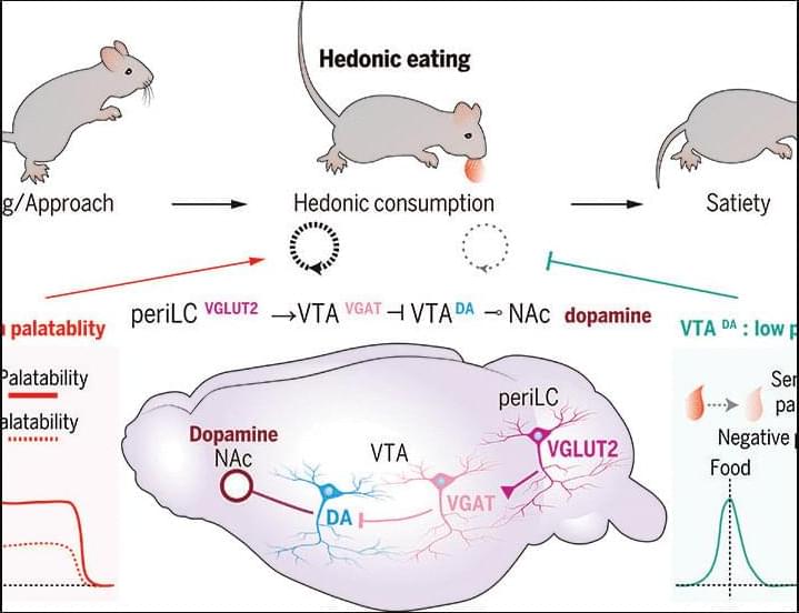
Hedonic eating is controlled by dopamine neurons that oppose GLP-1R satiety
Hedonic eating is defined as food consumption driven by palatability without physiological need. However, neural control of palatable food intake is poorly understood. We discovered that hedonic eating is controlled by a neural pathway from the peri–locus ceruleus to the ventral tegmental area (VTA). Using photometry-calibrated optogenetics, we found that VTA dopamine (VTADA) neurons encode palatability to bidirectionally regulate hedonic food consumption. VTADA neuron responsiveness was suppressed during food consumption by semaglutide, a glucagon-like peptide receptor 1 (GLP-1R) agonist used as an antiobesity drug. Mice recovered palatable food appetite and VTADA neuron activity during repeated semaglutide treatment, which was reversed by consumption-triggered VTADA neuron inhibition.
Does Methylene Blue Impact Lifespan?
Join us on Patreon! https://www.patreon.com/MichaelLustgartenPhD
Discount Links/Affiliates:
Blood testing (where I get the majority of my labs): https://www.ultalabtests.com/partners/michaellustgarten.
At-Home Metabolomics: https://www.iollo.com?ref=michael-lustgarten.
Use Code: CONQUERAGING At Checkout.
Clearly Filtered Water Filter: https://get.aspr.app/SHoPY
Epigenetic, Telomere Testing: https://trudiagnostic.com/?irclickid=U-s3Ii2r7xyIU-LSYLyQdQ6…M0&irgwc=1
Use Code: CONQUERAGING
NAD+ Quantification: https://www.jinfiniti.com/intracellular-nad-test/
Shocking Result from DESI Stuns Cosmology
Evolving cosmological constant.
Thanks to Storyblocks for sponsoring this video! Download unlimited stock media at one set price with Storyblocks: https://storyblocks.com/CoolWorlds.
The Standard Model of Cosmology has reigned supreme for decades, confirmed over and over again. But chinks in the armour have been developing, such as the Hubble Tension. Now, however, a new result threatens to completely upend our view of the Universe — we no longer even know how it will end.
Written & presented by Prof. David Kipping.
→ Support our research: https://www.coolworldslab.com/support.
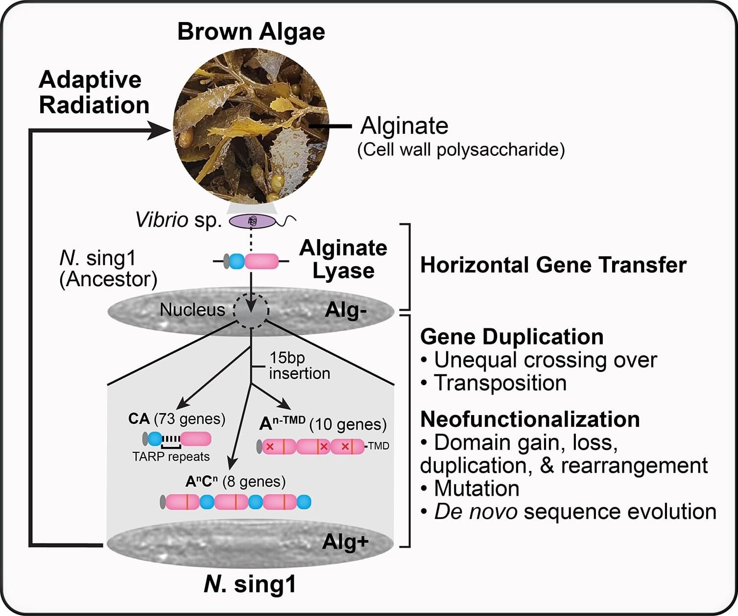
Giving up on photosynthesis: How a borrowed bacterial gene allows some marine diatoms to live on a seaweed diet
A group of diatom species belonging to the Nitzschia genus gave up on photosynthesis and now get their carbon straight from their environment, thanks to a bacterial gene picked up by an ancestor. Gregory Jedd of Temasek Life Sciences Laboratory, Singapore, and colleagues report these findings in a study published in the open-access journal PLOS Biology.
Unlike most diatoms, which perform photosynthesis to generate carbon compounds, some members of the genus Nitzschia have no chlorophyll and instead consume carbohydrates from seaweed and decaying plant matter. Previously, it was unclear how exactly they made this major lifestyle transition, so researchers sequenced the genome of one species, Nitzschia sing1, to look for clues.
N. sing1’s genome sequence showed that the diatom carries a gene for an enzyme that breaks down alginate, a carbon polymer in the cell walls of brown algae—a group that includes kelp and other common seaweeds. The gene originally came from a marine bacterium, and an ancestor of N. sing1 took up the gene and incorporated it into its genome.
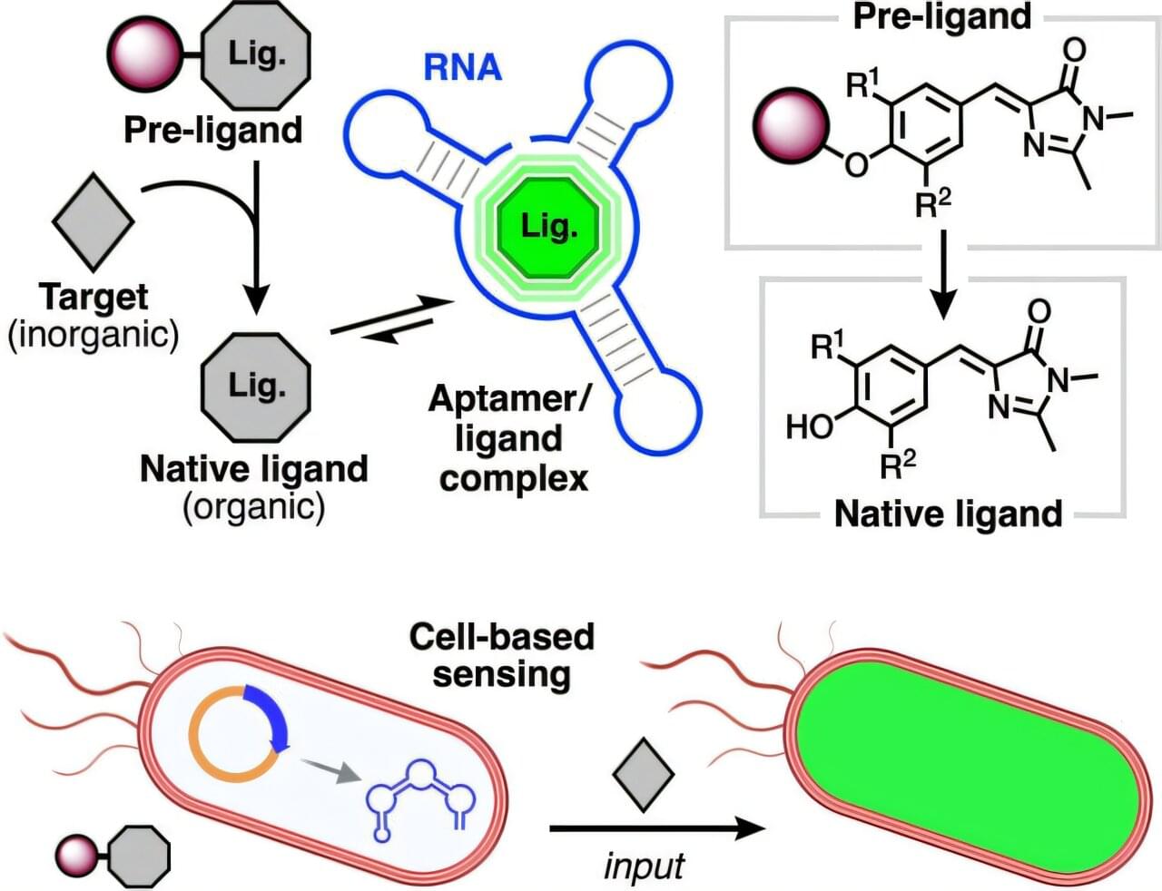
RNA transformed into biosensor for detecting health-related chemicals
Scientists have transformed RNA, a biological molecule present in all living cells, into a biosensor that can detect tiny chemicals relevant to human health.
Research by Rutgers University-New Brunswick scientists centers on RNA, a nucleic acid that plays a crucial role in most cellular processes. Their work is expected to have applications in the surveillance of environmental chemicals and, ultimately, the diagnosis of critical diseases including neurological and cardiovascular diseases and cancer.
“Imagine that people will go to the hospital and give a sample of cells from their own bodies for regular check-ups,” said Enver Cagri Izgu, an assistant professor in the Department of Chemistry and Chemical Biology in the Rutgers School of Arts and Sciences and the corresponding author of the study.
Scientists issue dire warning: Microplastic accumulation in human brains escalating
A new study published in Nature Medicine has revealed the presence of microplastics – tiny fragments of degraded plastic – in human brain tissue. While previous research has identified microplastics in organs such as the liver, kidneys, and placenta, this study suggests that the brain may be especially vulnerable to these tiny synthetic particles.
Scientists have made a disturbing discovery: human brains contain microplastics, and at higher concentrations than other organs. Worse, brain levels have jumped 50% in just eight years.