Human brains outperform computers in many forms of processing and are far more energy efficient. What if we could harness their power in a new form of biological computing?
In this Frontiers Forum Deep Dive session on 21 June 2023, Professor Thomas Hartung, Dr Lena Smirnova and other renowned researchers, explored the future of organoid intelligence and the scientific, technological and ethical steps required for realizing its full potential.
The session brought together the authors of the Frontiers in Science lead article ‘Organoid intelligence (OI): the new frontier in biocomputing and intelligence-in-a-dish’ which presents a roadmap for the strategic development of organoid intelligence as a scientific discipline. It was attended by hundreds of representatives from science, policy, and business across the world.
In multicellular organisms, many biological pathways exhibit a curious structure, involving sets of protein variants that bind or interact with one another in a many-to-many fashion. What functions do these seemingly complicated architectures provide? And can similar architectures be useful in synthetic biology? Here, Dr. Elowitz discusses recent work in his lab that shows how many-to-many circuits can function as versatile computational devices, explore the roles these computations play in natural biological contexts, and show how many-to-many architectures can be used to design synthetic multicellular behaviors.
About Michael Elowitz. Michael Elowitz is a Howard Hughes Medical Institute Investigator and Roscoe Gilkey Dickinson Professor of Biology and Biological Engineering at Caltech. Dr. Elowitz’s laboratory has introduced synthetic biology approaches to build and understand genetic circuits in living cells and tissues. As a graduate student with Stanislas Leibler, Elowitz developed the Repressilator, an artificial genetic clock that generates gene expression oscillations in individual E. coli cells. Since then, his lab has continued to design and build synthetic genetic circuits, bringing a “build to understand” approach to bacteria, yeast, and mammalian cells. He and his group have shown that gene expression is intrinsically stochastic, or ‘noisy’, and revealed how noise functions to enable probabilistic differentiation, time-based regulation, and other functions. Currently, Elowitz’s lab is bringing synthetic approaches to understand and program multicellular functions including multistability, cell-cell communication, epigenetic memory, and cell fate control, and to provide foundations for using biological circuits as therapeutic devices. His lab also co-develops systems such as “MEMOIR” that allows cells to record their own lineage histories and tools for RNA export, and precise gene expression. Elowitz received his PhD in Physics from Princeton University and did postdoctoral research at Rockefeller University. Honors include the HFSP Nakasone Award, MacArthur Fellowship, Presidential Early Career Award, Allen Distinguished Investigator Award, the American Academy of Arts and Sciences, and election to the National Academy of Sciences.
The Monthly Seminar on Physical Genomics is a public lecture series sponsored by the Center for Physical Genomics at Northwestern University, the Robert H. Lurie Comprehensive Cancer Center, and NIH Grants T32GM142604 and U54CA268084.
Summary: Researchers have discovered how glial cells can be reprogrammed into neurons through epigenetic modifications, offering hope for treating neurological disorders. This reprogramming involves complex molecular mechanisms, including the transcription factor Neurogenin2 and the newly identified protein YingYang1, which opens chromatin for reprogramming.
The study reveals how coordinated epigenome changes drive this process, potentially leading to new therapies for brain injury and neurodegenerative diseases.
Researchers have significantly improved gene-editing techniques. This new method, called eePASSIGE, can insert or replace entire genes in human cells with much higher efficiency than previous methods. This advancement could lead to a single gene therapy for diseases caused by various mutations in a single gene, like cystic fibrosis. Traditionally, gene therapy required a different treatment for each mutation.
EePASSIGE combines prime editing, which edits small stretches of DNA, with new enzymes that insert large pieces of DNA. This allows scientists to introduce a healthy copy of a gene directly where it belongs in the genome.
“This is one of the first examples of targeted gene integration with potential for therapeutic applications,” said Dr. David Liu, senior author of the study. “If these efficiencies translate to patients, many genetic diseases could be treated.”
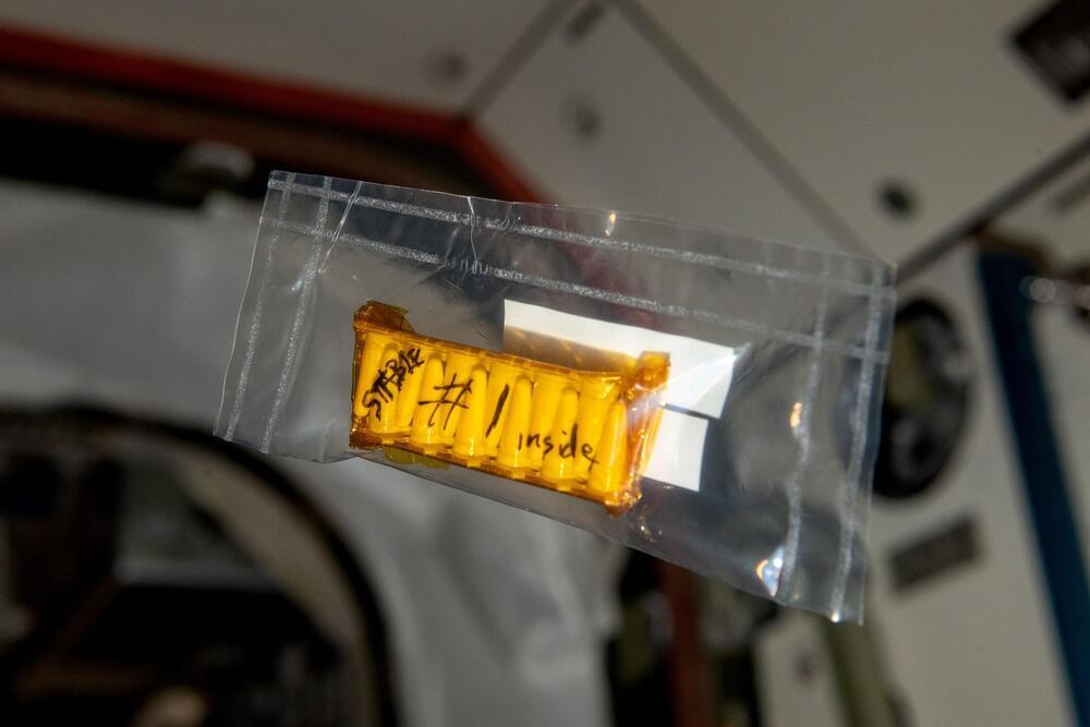
Microbes that are used for health, agricultural, or other applications need to be able to withstand extreme conditions, and ideally the manufacturing processes used to make tablets for long-term storage. MIT researchers have now developed a new way to make microbes hardy enough to withstand these extreme conditions.
Their method involves mixing bacteria with food and drug additives from a list of compounds that the FDA classifies as “generally regarded as safe.” The researchers identified formulations that help to stabilize several different types of microbes, including yeast and bacteria, and they showed that these formulations could withstand high temperatures, radiation, and industrial processing that can damage unprotected microbes.
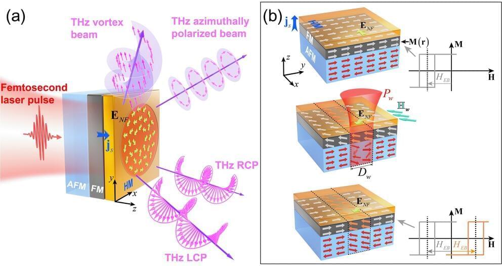
Researchers have developed a novel method for generating structured terahertz light beams using programmable spintronic emitters. This breakthrough offers a significant leap forward in terahertz technology, enabling the generation and manipulation of light with both spin and orbital angular momentum at these frequencies for the first time.
Terahertz radiation lies between microwaves and infrared light on the electromagnetic spectrum. It holds great promise for various applications, including security scanners, medical imaging, and ultrafast communication. However, generating and controlling terahertz light effectively has proven challenging.
This new research, published in eLight and led by Prof. Zhensheng Tao, Prof. Yizheng Wu from Fudan University and Prof. Yan Zhang from Capital Normal University, overcomes these limitations by employing programmable spintronic emitters based on exchange-biased magnetic multilayers. These devices consist of thin layers of magnetic and non-magnetic materials that convert laser-induced spin-polarized currents into broadband terahertz radiation.
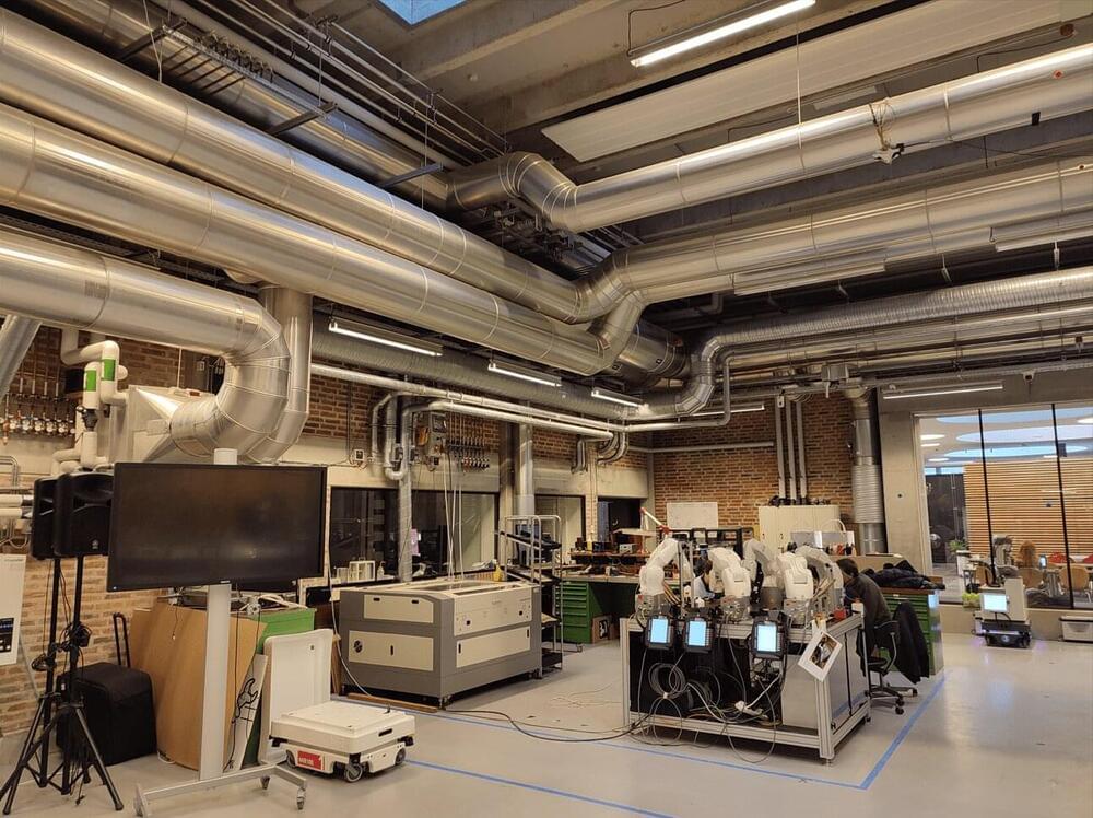
Researchers from the Smart and Wireless Applications and Technologies Group (SWAT-UGR) have conducted two scientific studies aimed at answering a common question: understanding how electromagnetic waves propagate in the medium.
The increase in network speed opens the door to new possibilities, such as robotic surgery or virtual reality services.
A team of UGR researchers has examined the propagation of electromagnetic waves with the goal of enhancing the deployment of 5G and 6G networks. Additionally, the study results contribute to the development of Industry 4.0, which seeks to automate processes in factories using wireless technologies.
Researchers have developed a pH-responsive nanorobot system that changes confirmation in the tumor microenvironment to selectively kill cancer cells in mice.
Researchers at the Karolinska Institutet (Stockholm, Sweden) have recently developed a nanorobot system capable of killing cancer cells in mice. This system works by activating at lower pH, such as within the tumor microenvironment. It is hoped that this could serve as a proof-of-concept for similar stimulus-responsive nanorobotic approaches and introduce a new range of effective cancer therapeutics.
Certain membrane proteins capable of inducing apoptosis, a type of cell death, appear on the surface of both healthy and cancer cells. These proteins, often called death receptors, join and activate when in close proximity to each other. This closeness is induced by external factors binding to the cell surface.
Consciousness is comprised of arousal (i.e., wakefulness) and awareness. Substantial progress has been made in mapping the cortical networks that modulate awareness in the human brain, but knowledge about the subcortical networks that sustain arousal is lacking. We integrated data from ex vivo diffusion MRI, immunohistochemistry, and in vivo 7 Tesla functional MRI to map the connectivity of a subcortical arousal network that we postulate sustains wakefulness in the resting, conscious human brain, analogous to the cortical default mode network (DMN) that is believed to sustain self-awareness. We identified nodes of the proposed default ascending arousal network (dAAN) in the brainstem, hypothalamus, thalamus, and basal forebrain by correlating ex vivo diffusion MRI with immunohistochemistry in three human brain specimens from neurologically normal individuals scanned at 600–750 µm resolution. We performed deterministic and probabilistic tractography analyses of the diffusion MRI data to map dAAN intra-network connections and dAAN-DMN internetwork connections. Using a newly developed network-based autopsy of the human brain that integrates ex vivo MRI and histopathology, we identified projection, association, and commissural pathways linking dAAN nodes with one another and with cortical DMN nodes, providing a structural architecture for the integration of arousal and awareness in human consciousness. We release the ex vivo diffusion MRI data, corresponding immunohistochemistry data, network-based autopsy methods, and a new brainstem dAAN atlas to support efforts to map the connectivity of human consciousness.
One sentence summary We performed ex vivo diffusion MRI, immunohistochemistry, and in vivo 7 Tesla functional MRI to map brainstem connections that sustain wakefulness in human consciousness.
BF has a financial interest in CorticoMetrics, a company whose medical pursuits focus on brain imaging and measurement technologies. BF’s interests were reviewed and are managed by Massachusetts General Hospital and Mass General Brigham HealthCare in accordance with their conflict-of-interest policies.
Learn more about the Cognitive Science Student Association and the California Cognitive Science Conference at https://cssa.berkeley.edu.
Amy Arnsten — Yale University.
Abstract. The recently evolved prefrontal cortex (PFC) subserves many of our highest-order cognitive functions, generating and sustaining the mental representations that underlie working memory, abstract reasoning, and top-down control of thought, action, and emotion. Due to the pioneering research of Patricia Goldman-Rakic, we have learned much about the neural basis underlying the ability of the dorsolateral prefrontal cortex (dlPFC) to generate mental representations, where microcircuits in deep layer III have extensive recurrent excitatory connections to maintain neuronal firing in the absence of sensory stimulation, while GABAergic interneurons provide lateral inhibition to refine the contents of working memory. However, these dlPFC circuits are also tremendously dependent on arousal state, with a narrow inverted U response to levels of acetylcholine, dopamine and norepinephrine. Even quite mild uncontrollable stress increases the release of dopamine and norepinephrine in the PFC, which rapidly impairs PFC functioning by 1) stimulating D1 and alpha-1-receptors, respectively, 2) these, in turn, activate feedforward calcium-cAMP signaling within spines, which then 3) open nearby potassium channels to disconnect PFC networks and take the PFC “off-line”. With chronic stress exposure, there is actual atrophy of PFC dendrites and spines. Understanding the neural events that weaken vs. strengthen PFC connectivity and function has led to the development of treatments for patients with stress-related PFC dysfunction, e.g. guanfacine and prazosin. This knowledge is also helping to illuminate the etiology of cognitive disorders, as genetic insults in schizophrenia often increase the activity of these stress signaling pathways, and the molecules that regulate the stress signaling pathways are lost with advancing age, leading to tau pathology as seen in Alzheimer’s disease.