As we turn our attention from the front of the eye to the back, we also look to the future. Many studies have combined oculomics with AI tools to predict biological age from retinal biomarkers, such as retinal vasculature [1, 6], and even linked this to chronic disease risk, such as cardiovascular disease and cancer [7]. High resolution imaging tools also enable direct visualisation of the neural layers within the retina, which can show signs of neurodegenerative diseases, such as Alzheimer’s disease [1, 6], Parkinson’s disease [8], multiple sclerosis [6, 9], and even rare conditions, such as Lafora disease [10]. In many cases, the oculomic signs are present before symptoms arise. For example, it has been shown that proteins related to Alzheimer’s disease (such as amyloid-beta) accumulate at least one decade prior to cognitive decline [11] and these proteins also accumulate in the retina [12]. This is particularly pertinent to clinical research and drug development, as it enables identification of those who may benefit from intervention before irreversible damage has taken place.
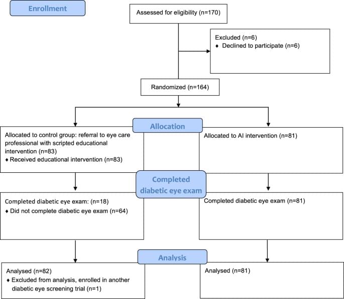
Advances in imaging technology mean that we can now detect biomarkers at cellular resolution. We are continually finding new applications for imaging techniques to detect disease before it takes hold, providing the opportunity to intervene and potentially avoid disease altogether. It’s definitely an exciting time for oculomics research!
Crystallomancy has come a long way since Ancient Roman times, and it makes one wonder whether the scryers of the past could have predicted the transformation of orb-gazing from a mystical art to a rigorous science. Not only does Oculomics enable us to look into your past and present, but also has the potential to look into your future, providing you the opportunity to change your “fate”. Although we cannot be sure what form the advancements in imaging and AI tools will take over the coming years, we can be sure of one thing – that oculomics has a promising future in the quest for longevity.