Feb 25, 2023
Cardiovascular Complications of Viral Respiratory Infections and COVID-19
Posted by Shubham Ghosh Roy in category: biotech/medical
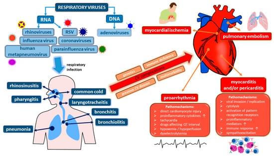
Viral respiratory infections (VRI) are the most prevalent type of infectious diseases and constitute one of the most common causes of contact with medical care. Regarding the pathophysiology of the cardiovascular system, VRI can not only exacerbate already existing chronic cardiovascular disease (such as coronary artery disease or heart failure) but also trigger new adverse events or complications (e.g., venous thromboembolism), the latter particularly in subjects with multimorbidity or disease-related immobilization. In the current paper, we provide a narrative review of diverse cardiovascular complications of VRI as well as summarize available data on the pathology of the circulatory system in the course of coronavirus disease 2019 (COVID-19).