Toggle light / dark theme

Just published from my son.

Automatic hippocampus imaging, with about 20 minutes of cloud computing per scan.


Like neocortical structures, the archicortical hippocampus differs in its folding patterns across individuals. Here, we present an automated and robust BIDS-App, HippUnfold, for defining and indexing individual-specific hippocampal folding in MRI, analogous to popular tools used in neocortical reconstruction. Such tailoring is critical for inter-individual alignment, with topology serving as the basis for homology. This topological framework enables qualitatively new analyses of morphological and laminar structure in the hippocampus or its subfields. It is critical for refining current neuroimaging analyses at a meso-as well as micro-scale. HippUnfold uses state-of-the-art deep learning combined with previously developed topological constraints to generate uniquely folded surfaces to fit a given subject’s hippocampal conformation. It is designed to work with commonly employed sub-millimetric MRI acquisitions, with possible extension to microscopic resolution. In this paper, we describe the power of HippUnfold in feature extraction, and highlight its unique value compared to several extant hippocampal subfield analysis methods.

Keywords: Brain Imaging Data Standards; computational anatomy; deep learning; hippocampal subfields; hippocampus; human; image segmentation; magnetic resonance imaging; neuroscience.

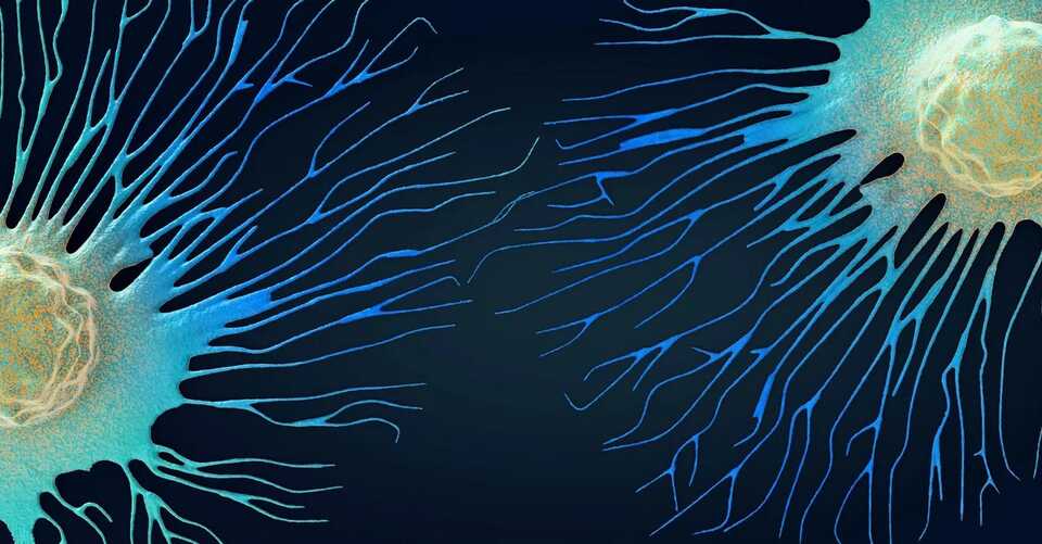
https://img.particlenews.com/image.php?url=2Ghcdg_0kAkXsy000 Tumor cells traverse many different types of fluids as they travel through the body. Christoph Burgstedt/Science Photo Library via Getty Images.
This article was originally featured on The Conversation.

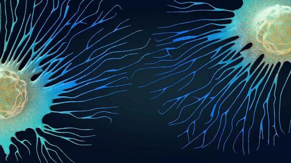
Cell migration, or how cells move in the body, is essential to both normal body function and disease progression. Cell movement is what allows body parts to grow in the right place during early development, wounds to heal and tumors to become metastatic.

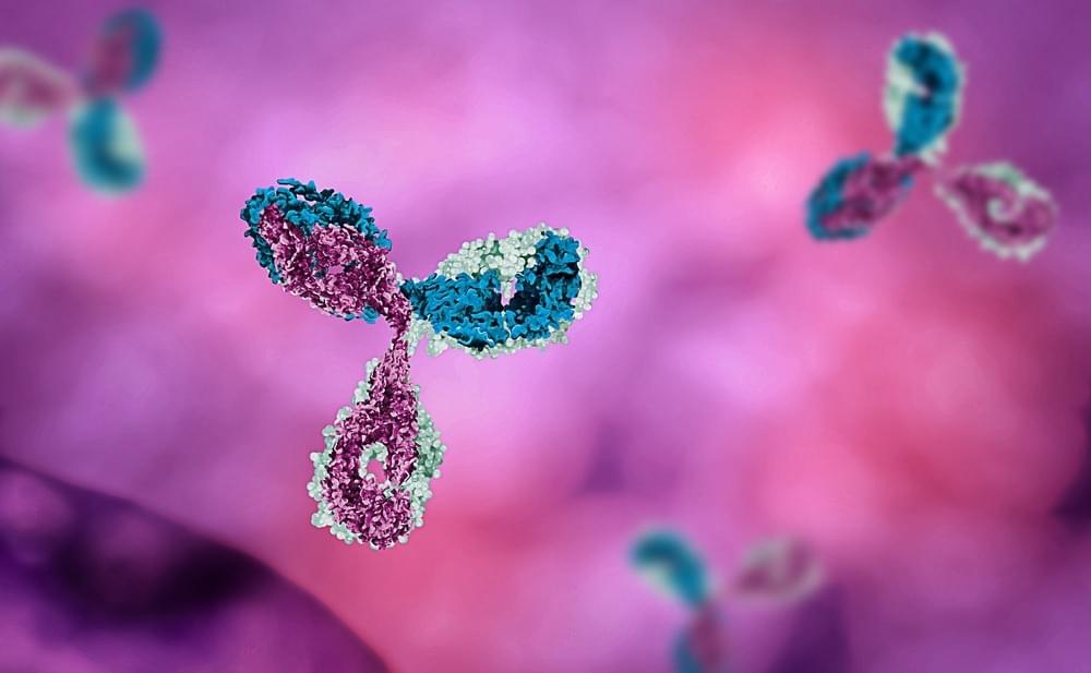
Even though the clinical efficacy of antibody-based therapeutics has been established, no methods that involve the de novo design of antibodies with wet lab validation are available.

About the study

A recent study, posted in the bioRxiv* preprint server, used generative AI models to develop de novo design antibodies against three distinct targets in a zero-shot fashion. A zero-shot designing method involves designing an antibody to bind to an antigen without follow-up optimization. The newly designed process has been termed de novo, meaning proteins (antibodies) were designed from first principles or from scratch.

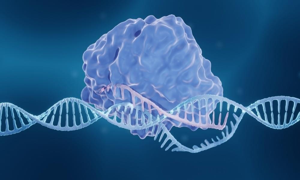
In this interview, News Medical speaks to Assistant Professor Ryan Jackson about his latest work, published in tandem Nature papers, detailing the discovery of a new CRISPR immune system.

Please can you introduce yourself and tell us about your professional background?

I am an Assistant Professor at Utah State University (USU). I use biochemical and structural techniques to understand how the molecules that perform the reactions of life function. I’ve been working in the CRISPR field since 2011. I started as a postdoc in Blake Wiedenheft’s lab at Montana State University, and in 2016 I started my own research lab at USU. I earned both of my degrees (a B.S. in Biology and a Ph.D. in Biochemistry) from USU, so joining the faculty was like coming home. My research lab specializes in determining the structure and function of newly discovered and obscure CRISPR systems.

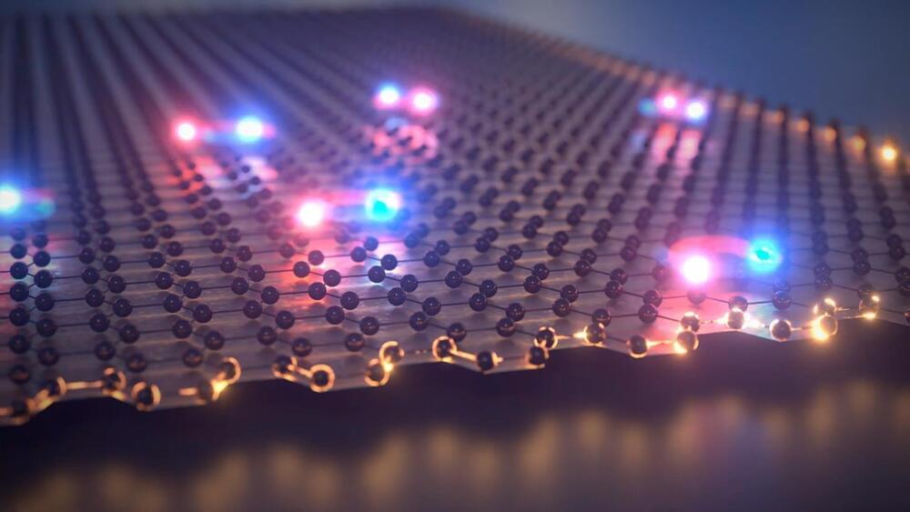
A breakthrough in quantum research – the first detection of excitons (electrically neutral quasiparticles) in a topological insulator has been achieved by an international team of scientists collaborating within the Würzburg-Dresden Cluster of Excellence ct.qmat. This discovery paves the way for a new generation of light-driven computer chips and quantum technologies. It was enabled thanks to smart material design in Würzburg, the birthplace of topological insulators. The findings have been published in the journal Nature Communications.

<em>Nature Communications</em> is a peer-reviewed, open-access, multidisciplinary, scientific journal published by Nature Portfolio. It covers the natural sciences, including physics, biology, chemistry, medicine, and earth sciences. It began publishing in 2010 and has editorial offices in London, Berlin, New York City, and Shanghai.

A comprehensive analysis of the cryptographic protocols used in the Swiss encrypted messaging application Threema has revealed a number of loopholes that could be exploited to break authentication protections and even recover users’ private keys.

The seven attacks span three different threat models, according to ETH Zurich researchers Kenneth G. Paterson, Matteo Scarlata, and Kien Tuong Truong, who reported the issues to Threema on October 3, 2022. The weaknesses have since been addressed as part of updates released by the company on November 29, 2022.

Threema is an encrypted messaging app that’s used by more than 11 million users as of October 2022. “Security and privacy are deeply ingrained in Threema’s DNA,” the company claims on its website.

Dr Vittorio Sebastiano presents about aging and reprogramming and answers questions from audience in this clip. He specifies short Reprogramming does not impact cellular Identity but Impact cellular age and cellular health.

Dr. Vittorio Sebastiano is an Assistant Professor in the Department of Obstetrics and Gynecology at Stanford School of Medicine. His lab has established a new technology named ERA (Epigenetic Reprogramming of Aging), which repurposes the conceptual idea of reprogramming, with the goal to promote epigenetic rejuvenation of adult cells leaving their identity untouched. This new technology was patented and is being implemented by Turn Biotechnologies, of which Dr. Sebastiano is co-founder and Chair of the Scientific Advisory Board.

In 2009, Dr. Sebastiano completed a postdoctoral fellowship at the laboratory of Dr. Marius Wernig at Stanford University, where he implemented the newly discovered iPSC technology and was among the first to demonstrate that iPSCs can be efficiently derived, genetically modified, and implemented for cell therapy in genetic diseases (Sebastiano et al., 2014, Science Translational Medicine).
Dr. Sebastiano completed his undergraduate and graduate studies at the University of Pavia, Italy, where he studied murine germ cells and preimplantation development and where he pioneered cellular reprogramming by Somatic Cell Nuclear Transfer. He joined the Max Planck Institute for Molecular Biomedicine as a postdoctoral fellow under the mentorship of Dr. Hans Robert Schöler, where he continued his research on cellular reprograming, germ cells biology, and embryonic development.

DISCLAIMER: Please note that none of the information in this video constitutes health advice or should be substituted in lieu of professional guidance. The video content is purely for informational purposes.

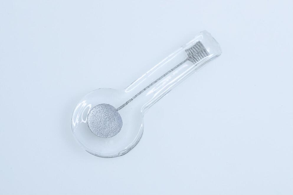
Hydrogels are three-dimensional (3D) polymer networks that do not dissolve in water but retain large amounts of liquids. Due to this advantageous property, hydrogels are particularly promising material platforms for both biomedical and environmental applications, as they can survive in bodily fluids or in wet natural environments without dissipating.

Over the past decade, engineers and materials scientists have been developing numerous based on soft hydrogels, including environmental and biomedical sensors, drug delivery devices, and artificial tissue. Despite the huge potential of these -based devices, their widespread implementation has so far been hindered by their high production costs.

A research team led by Dr. Nanjia Zhou at Westlake University and Westlake Institute of Advanced Studies in China have recently introduced a new strategy to enable the 3D printing of soft hydrogel electronics. Their approach, introduced in a paper published in Nature Electronics, could help to lower the production costs of numerous hydrogel-based devices, including strain sensors, inductors, and biological electrodes.

New tools are steadily bridging this gap. And ongoing development of one particular technique, cryo-electron tomography, or cryo-ET, has the potential to deepen how researchers study and understand how cells function in health and disease.

As the former editor-in-chief of Science magazine and as a researcher who has studied hard-to-visualize large protein structures for decades, I have witnessed astounding progress in the development of tools that can determine biological structures in detail. Just as it becomes easier to understand how complicated systems work when you know what they look like, understanding how biological structures fit together in a cell is key to understanding how organisms function.