Toggle light / dark theme

A frightening virus is killing a massive number of wild birds

Remember when eggs were so high? A vaccine for birds, now that can make money. đŸ€”

In the past two years, a viral disease has swept across much of the planet — not Covid but a type of avian flu. It’s devastated the poultry industry in the US, Europe, and elsewhere, sickening millions of farmed birds, which either die from infection or are killed by farmers seeking to stem the spread.


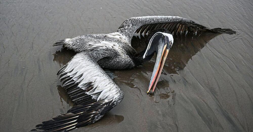
The ongoing outbreak of avian flu has killed hundreds of thousands — if not millions — of wild birds, including endangered species like the California condor. It’s one of the worst wildlife disease outbreaks in history. Having now spread across five continents and hundreds of wildlife species, scientists call the current outbreak a panzootic, meaning a pandemic among animals.

“What we’re seeing right now is uncharted territory,” said Andrew Ramey, a wildlife geneticist at the US Geological Survey, one of the federal agencies involved in testing wild birds for the virus.

The number of dead birds in itself is historic, but so is the virus’s biology. Typically, avian influenza viruses only cause severe disease and death in domestic birds like chickens and farmed ducks; they sweep through populations, killing upward of 90 percent of the flock.

Extracellular Vesicles from Stem Cells Reverse Senescence

Scientists have found that bubbles secreted by embryonic stem cells counter cellular senescence, in large part due to just two tiny snippets of RNA [1].

Extracellular vesicles (EVs) are membrane-bound tiny bubbles that are loaded with various molecular cargo, such as proteins, DNA, or RNA, that cells secrete as a method of intercellular communication. According to numerous studies, EVs can recapitulate many effects of cellular therapies, such as stem cell treatments [2].

In this new study, the researchers used EVs derived from human embryonic stem cells (ESC) against cellular senescence. ESCs are considered a potent therapeutic, tool but, as the study’s authors note in the introduction, their use “is limited by immune rejection, tumorigenicity and ethical issues”. If we could culture ESCs and use EVs secreted by them to the same effect, this would solve many problems.

Researchers achieve high-speed super-resolution imaging with a large field of view

Researchers have developed a fluorescence microscope that uses structured illumination for fast super-resolution imaging over a wide field of view. The new microscope was designed to image multiple living cells simultaneously with a very high resolution to study the effects of various drugs and mixtures of drugs on the body.

“Polypharmacy—the effect of the many combinations of drugs typically prescribed to the chronically sick or elderly—can lead to dangerous interactions and is becoming a major issue,” said Henning Ortkrass from Bielefeld University in Germany. “We developed this microscope as part of the EIC Pathfinder OpenProject DeLIVERy, which aims to develop a platform that can investigate polypharmacy in individual patients.”

In the journal Optics Express, the researchers describe their new microscope, which uses optical fiber delivery of excitation light to enable very high image quality over a very large field of view with multicolor and high-speed capability. They show that the instrument can be used to image , achieving a field of view up to 150 × 150 ÎŒm2 and imaging rates up to 44 Hz while maintaining a spatiotemporal resolution of less than 100 nm.

No More Cavities? Organoids Pave the Way for Enamel Regeneration

Stem cells have been used to produce organoids that release the proteins responsible for forming dental enamel, a substance that shields teeth from harm and decay. This initiative was led by a multi-disciplinary team of researchers from the University of Washington in Seattle.

“This is a critical first step to our long-term goal to develop stem cell-based treatments to repair damaged teeth and regenerate those that are lost,” said Hai Zhang, professor of restorative dentistry at the UW School of Dentistry and one of the co – authors of the paper describing the research.

The findings are published today in the journal Developmental Cell. Ammar Alghadeer, a graduate student in Hannele Ruohola-Baker’s laboratory in the Department of Biochemistry at the UW School of Medicine was the lead author on the paper. The lab is affiliated with the UW Medicine Institute for Stem Cell and Regenerative Medicine.

New insights into human neurogenesis: Researchers discover key role of amyloid precursor protein

A recent study study sheds light on how a protein called amyloid precursor protein (APP) affects the growth of nerve cells in the cortex — the human brain’s outer layer. The findings suggest that APP plays a crucial role in maintaining the delicate balance between neural stem cell proliferation and differentiation during the early stages of brain development.

The research, published in Science Advances, could have important implications for our understanding of neurodevelopmental processes and neurodegenerative diseases.

APP is a class I transmembrane protein that is widely expressed during nervous system development. It has been extensively studied due to its connection to Alzheimer’s disease (AD), where its fragmentation produces amyloid peptides that contribute to neuronal death. However, the physiological function of APP, especially in the context of human brain development, has remained unclear.

Brain imaging study suggests that loss of consciousness is related to the malfunctioning of two neural circuits

A team of scientists recently aimed to better understand consciousness and its pathologies by studying the neural activity of patients with disorders of consciousness and healthy volunteers using brain imaging technology. They identified two crucial brain circuits implicated in consciousness. The results of the study have been published in Human Brain Mapping.

Consciousness is a complex and subjective experience, and there is still much debate among scientists and philosophers about its nature and origin. However, in clinical settings, doctors treating patients with severe brain injuries and disorders of consciousness need to find ways to help their patients, regardless of the exact definition of consciousness. The authors of the new study sought to better understand the mechanisms behind the pathological loss of consciousness and its recovery, as well as to have reliable ways to assess the state of the patients.

“In recent years, many studies have tried to objectively assess levels of consciousness using various neuroimaging techniques. While these studies have improved how we diagnose patients with disorders of consciousness, they haven’t fully explained how consciousness comes about,” explained study author Jitka Annen, a postdoctoral researcher at the Coma Science Group at the University of Liege.

Neuroscientists Re-create Pink Floyd Song from Listeners’ Brain Activity

Neuroscientists have worked for decades to decode what people are seeing, hearing or thinking from brain activity alone. In 2012 a team that included the new study’s senior author—cognitive neuroscientist Robert Knight of the University of California, Berkeley—became the first to successfully reconstruct audio recordings of words participants heard while wearing implanted electrodes. Others have since used similar techniques to reproduce recently viewed or imagined pictures from participants’ brain scans, including human faces and landscape photographs. But the recent PLOS Biology paper by Knight and his colleagues is the first to suggest that scientists can eavesdrop on the brain to synthesize music.

“These exciting findings build on previous work to reconstruct plain speech from brain activity,” says Shailee Jain, a neuroscientist at the University of California, San Francisco, who was not involved in the new study. “Now we’re able to really dig into the brain to unearth the sustenance of sound.”

To turn brain activity data into musical sound in the study, the researchers trained an artificial intelligence model to decipher data captured from thousands of electrodes that were attached to the participants as they listened to the Pink Floyd song while undergoing surgery.

Department of Energy-Funded Quantum Rods Breakthrough Could Enable Ultra High-Def Virtual Reality

A breakthrough at the Massachusetts Institute of Technology (MIT), funded by the Department of Energy (DOE), is utilizing the power of DNA to align quantum rods that may help usher in a new era of enhanced televisions and ultra-realistic virtual reality (VR) devices.

Some modern high-end TVs already make use of the dynamic luminescent qualities of two-dimensional (2D) quantum dots, but finding a way to tap into the superior qualities of their two-dimensional cousins, quantum rods, has remained elusive. However, the researchers behind this latest effort claim that the situation has now changed.

The Secrets of Realistic VR and Star Trek Holodecks May Lie in the Quantum Realm.