Toggle light / dark theme

Astrocytes: a hub of olfactory sensation processing

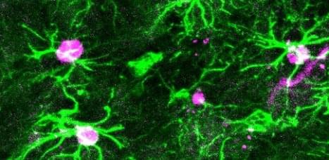
To enjoy the scent of morning coffee and freshly baked cookies or to perceive the warning smell of something burning, the brain needs two types of cells, neurons and astrocytes, to work closely with each other. Research has shown a great deal of the changes that occur in neurons during olfactory, or smell, perception, but what are the astrocyte responses and how they contribute to the sensory experience remains unclear.

Researchers at Baylor College of Medicine and collaborating institutions report in the journal Science the responses of astrocytes to olfactory stimulation, revealing a new mechanism that is required to maintain astrocyte-neuron communication and process olfactory sensation.

“Previous studies have shown that under natural conditions in a living animal, olfactory stimulation of the brain activates neurons first, which changes the genes these neurons express to be able to mediate the olfactory sensation,” said first author Dr. Debosmita Sardar, a postdoctoral associate in Dr. Benjamin Deneen’s lab at Baylor. “In this study, we investigated what occurred to astrocytes following neural activity during olfactory stimulation and uncovered changes that had not been described before.”

Epigenetic Factors Create the Immune System’s Memory

The immune system is an incredibly complex network that has some amazing capabilities. It can eliminate dangerous cells that may lead to cancer, and defend the body against a wide variety of pathogenic invaders. It also has the ability to remember those encounters with pathogens so if they happen again, the immune system is primed to respond more quickly and forcefully against the offender. Scientists have now learned more about how the immune system memory is created at the molecular level. The findings have been reported in Science Immunology.

When immune cells are exposed to an invader, they can recognize structures called antigens on the surface of the pathogen. In this study, the researchers compared immune cells that had never been exposed to an antigen, so-called naive cells, to immune cells that had been in contact with an antigen, known as memory cells. The investigators wanted to identify the epigenetic differences between these cell types, which are changes in DNA that can impact gene expression, such as structural shifts or chemical tags, but do not alter the sequence of the genome. Epigenetic changes might explain why memory cells can react so quickly while naive cells are comparatively slow.

This company wants you to live forever in their metaverse

Consider it a technological solution to the problem of death.

Over the last couple years, I’ve been writing about creating ghosts — perhaps an inevitability in the midst of a pandemic.

Artur Sychov, founder and CEO of metaverse company Somnium Space, has joined the quest against loss. Using motion capture and voice data, he wants to create duplicate avatars that can move as you moved and speak as you spoke, using your voice.

How to grow new brain cells

For a long time, neuroscientists believed that the neurons you are born with are the neurons you have for the rest of your life, and any neuron lost will not be replaced. Recent research has shown that specific brain regions contain neural stem cells that can generate new neurons. In this talk, Dr Daniel Berg of the University of Aberdeen will discuss what we know about these stem cells and what we can do to activate them to generate more neurons.

Fred Gage — Regulation and Function of Neurogenesis in the Adult Hippocampus

Watch this presentation on LabRoots at: http://www.labroots.com/webcast/keynote-speaker-regulation-a…ippocampus.

In the adult central nervous system (CNS) small populations of neurons are formed in the adult olfactory bulb and dentate gyrus of the hippocampus. In the adult hippocampus, newly born neurons originate from stem cells that exist in the subgranular zone of the dentate gyrus. Progeny of these putative stem cells differentiate into neurons in the granular layer within a month of the cells’ birth, and this late neurogenesis continues throughout the adult life of all mammals. Environmental stimulation can differentially effect the proliferation, migration and differentiation of these cells in vivo. These environmentally induced changes in the structural organization of the hippocampus, result in changes in electrophysiological responses in the hippocampus, as well as in hippocampal related behaviors. We are studying the cellular, molecular, as well as environmental influences that regulate neurogenesis in the adult brain. We have recently identified several molecules that work coordinately to regulate proliferation, survival and differentiation of these adult derived stem cells. In addition, we have demonstrated that specific types of activity can influence the behavior of these newly born cells. Finally, we have developed several methods to monitor the in vivo maturation of neurogenesis in vivo, which has provided insight to the functional importance of neurogenesis to behavior. A consensus model of the function of adult neurogenesis is emerging.

Research reveals Butterflies, moths share ancient ‘blocks’ of DNA

According to new research, butterflies and moths share “blocks” of DNA that are more than 200 million years old. Scientists from the Universities of Exeter (UK), Lübeck (Germany) and Iwate (Japan) devised a tool to compare the chromosomes (DNA molecules) of different butterflies and moths.

They found blocks of chromosomes that exist in all moth and butterfly species, and also in Trichoptera – aquatic caddisflies that shared a common ancestor with moths and butterflies some 230 million years ago. Moths and butterflies (collectively called Lepidoptera) have widely varying numbers of chromosomes – from 30 to 300 – but the study’s findings show remarkable evidence of shared blocks of homology (similar structure) going back through time.

“DNA is compacted into individual particles or chromosomes that form the basic units of inheritance,” said Professor Richard ffrench-Constant, from the Centre for Ecology and Conservation on Exeter’s Penryn Campus in Cornwall. “If genes are on the same ‘string’, or chromosome, they tend to be inherited together and are therefore ‘linked’.

Cellular deconvolution with continuous transitions

A recent work introduces a cellular deconvolution method, MeDuSA, of estimating cell-state abundance along a one-dimensional trajectory from bulk RNA-seq data with fine resolution and high accuracy, enabling the characterization of cell-state transition in various biological processes.

Single-cell transcriptomic techniques continue to revolutionize the resolution of cell analysis, determining discrete cell types and cell states with continuous dynamic transitions that can be related to development and disease progression5. Cells in different states can be computationally ordered according to a pseudo-time series, or cell trajectory6. Both MeDuSA and another method, Cell Population Mapping (CPM)7, were developed to exploit the rich spectrum of single-cell reference profiles to estimate cell-state abundance in bulk RNA-seq data, which enables fine-resolution cellular deconvolution (Fig. 1b). Although CPM effectively tackles the issue of estimating the abundance of cells in different states, MeDuSA further improves the estimation accuracy by employing a LMM (see the equation in Fig. 1c) that takes into account both the cell state of interest (focal state) and the remaining cells of the same cell type (non-focal state) as well as the other cell types.

OrganoidChip facilitates hydrogel-free immobilization for fast and blur-free imaging of organoids

To show the capability of the OrganoidChip in enabling higher-resolution imaging, we used confocal microscopy for several organoids immobilized on the chip. Representative images show improved optical segmentation and the ability to resolve single cells within an organoid (Fig. 4 d). The co-localized EthD-1-and Hoechst-stained nuclei are resolvable and can potentially be used to increase the accuracy of viability measurements. Future implementation of 3D-segmentation using AI-assisted algorithms in the analysis pipeline can provide more accurate estimations of cellular viability in larger screens.

Next, we measured the effect of DOX treatment on the beating kinetics of cardiac organoids. To do this, we relied on calcium fluorescence imaging, as it has been shown to be a good approximation of the cardiomyocytes’ action potentials32. Calcium imaging proved beneficial for beating and contraction parameters since smaller beating portions cannot necessarily be detected from brightfield images, particularly when organoids have been compromised as a result of drug treatment.

When assessing drug effects, we observed some degree of variability in the spontaneous contractile behaviour and beating kinetics between cardiac organoids. Such variability often skews any averaged parameter value across organoids and does not reflect the effect of the treatment conditions on organoid health. To address this challenge, we tracked each individual organoid’s beating off-and on-chip. The drug-induced functionality results are therefore reported as averages of fractional changes of each individual organoid’s beating kinetics parameters, measured at 48 h post-treatment, on both the chamber slide and on the chip, relative to its pre-treatment value (Eq. 3).