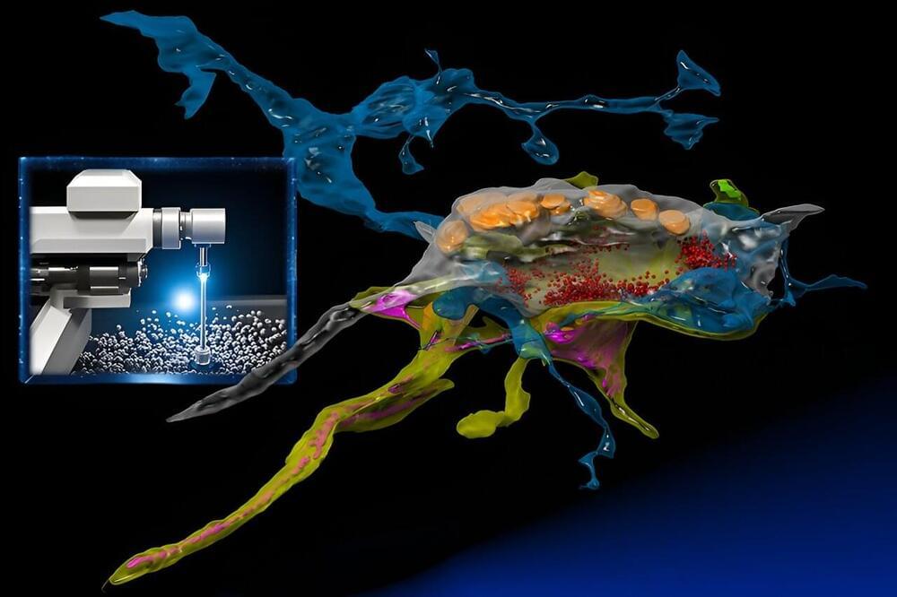
Connectomics, the ambitious field of study that seeks to map the intricate network of animal brains, is undergoing a growth spurt. Within the span of a decade, it has journeyed from its nascent stages to a discipline that is poised to (hopefully) unlock the enigmas of cognition and the physical underpinning of neuropathologies such as in Alzheimer’s disease.
At its forefront is the use of powerful electron microscopes, which researchers from the MIT Computer Science and Artificial Intelligence Laboratory (CSAIL) and the Samuel and Lichtman Labs of Harvard University bestowed with the analytical prowess of machine learning. Unlike traditional electron microscopy, the integrated AI serves as a “brain” that learns a specimen while acquiring the images, and intelligently focuses on the relevant pixels at nanoscale resolution similar to how animals inspect their worlds.
“SmartEM” assists connectomics in quickly examining and reconstructing the brain’s complex network of synapses and neurons with nanometer precision. Unlike traditional electron microscopy, its integrated AI opens new doors to understand the brain’s intricate architecture. “SmartEM: machine-learning guided electron microscopy” has been published on the pre-print server bioRxiv.


Comments are closed.