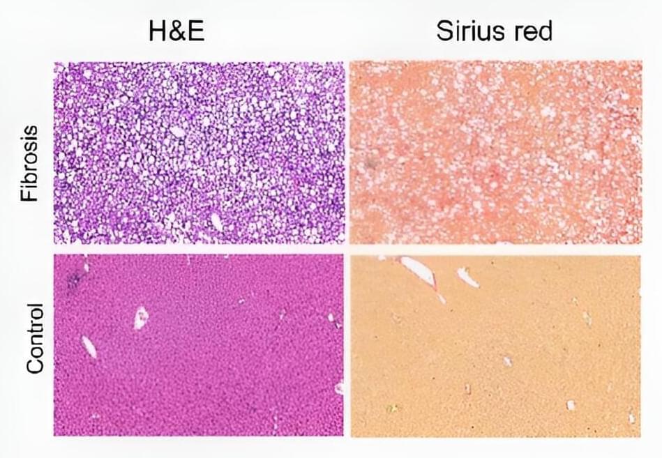
Chronic liver diseases such as hepatitis, liver cirrhosis, and hepatocellular carcinoma are a major cause of morbidity and mortality worldwide. Fibrosis—the thickening and scarring of connective tissue—plays a major role in these liver diseases but detection of fibrosis is limited to biopsy, which suffers from limitations including the risk of complications, sampling only a tiny fraction of the liver, and an inability to serially monitor disease progression due to its invasive nature.
To provide better diagnosis and treatment of chronic liver diseases, researchers are working to use non-invasive magnetic resonance imaging (MRI) to detect and quantify liver fibrosis throughout the entire organ, which would enable earlier detection and the ability to monitor disease progression as well as the effects of treatment over time.
Adapting MRI for detecting chronic conditions such as fibrosis involves the development of tissue-specific MRI contrast agents that target diseased tissue such as the collagen that accumulates in fibrotic liver. To develop such agents, researchers have been challenged to design and synthesize compounds that must find and bind the tissue target, provide a strong signal under MRI, and rapidly clear from the system to minimize any toxicity.


Comments are closed.