Toggle light / dark theme


Image copyright of Augusta University

Our brains have a basic algorithm that enables us to not just recognize a traditional Thanksgiving meal, but the intelligence to ponder the broader implications of a bountiful harvest as well as good family and friends.

“A relatively simple mathematical logic underlies our complex brain computations,” said Dr. Joe Z. Tsien, neuroscientist at the Medical College of Georgia at Augusta University, co-director of the Augusta University Brain and Behavior Discovery Institute and Georgia Research Alliance Eminent Scholar in Cognitive and Systems Neurobiology.

Read more

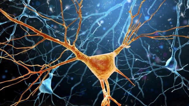
In a cross-domain study directed by professor Peter Carmeliet (VIB — KU Leuven), researchers discovered unexpected cells in the protective membranes that enclose the brain, the so called meninges. These ‘neural progenitors’ — or stem cells that differentiate into different kinds of neurons — are produced during embryonic development. These findings show that the neural progenitors found in the meninges produce new neurons after birth — highlighting the importance of meningeal tissue as well as these cells’ potential in the development of new therapies for brain damage or neurodegeneration. A paper highlighting the results was published in the leading scientific journal Cell Stem Cell.

Scientists’ understanding of brain plasticity, or the ability of the brain to grow, develop, recover from injuries and adapt to changing conditions throughout our lives, has been greatly broadened in recent years. Before the discoveries of the last few decades, neurologists once thought that the brain became ‘static’ after childhood. This dogma has changed, with researchers finding more and more evidence that the brain is capable of healing and regenerating in adulthood, thanks to the presence of stem cells. However, neuronal stem cells were generally believed to only reside within the brain tissue, not in the membranes surrounding it.

The meninges: unappreciated no more: Believed in the past to serve a mainly protective function to dampen mechanical shocks, the meninges have been historically underappreciated by science as having neurological importance in its own right. The data gathered by the team challenges the current idea that neural precursors — or stem cells that give rise to neurons — can only be found inside actual brain tissue.

Read more

BENGALURU: After working for five years, a team of three from department of Biosciences and Bioengineering (BSBE) at Indian Institute of Technology (IIT), Bombay and IITB-Monash Research Academy has designed smart amyloid based hydrogels that are able to guide stem cell to differentiate to neuron and successfully transplanted these stem cells in the brain of Parkinson’s disease (PD) animal models with unique amyloid hydrogels.

Read more

New biomarkers for aging is good news for researchers!


“Given the high volume of data being generated in the life sciences, there is a huge need for tools that make sense of that data. As such, this new method will have widespread applications in unraveling the molecular basis of age-related diseases and in revealing biomarkers that can be used in research and in clinical settings. In addition, tools that help reduce the complexity of biology and identify important players in disease processes are vital not only to better understand the underlying mechanisms of age-related disease but also to facilitate a personalized medicine approach. The future of medicine is in targeting diseases in a more specific and personalized fashion to improve clinical outcomes, and tools like iPANDA are essential for this emerging paradigm,” said João Pedro de Magalhães, PhD, a trustee of the Biogerontology Research Foundation.

The algorithm, iPANDA, applies deep learning algorithms to complex gene expression data sets and signal pathway activation data for the purposes of analysis and integration, and their proof of concept article demonstrates that the system is capable of significantly reducing noise and dimensionality of transcriptomic data sets and of identifying patient-specific pathway signatures associated with breast cancer patients that characterize their response to Toxicol-based neoadjuvant therapy.

The system represents a substantially new approach to the analysis of microarray data sets, especially as it pertains to data obtained from multiple sources, and appears to be more scalable and robust than other current approaches to the analysis of transcriptomic, metabolomic and signalomic data obtained from different sources. The system also has applications in rapid biomarker development and drug discovery, discrimination between distinct biological and clinical conditions, and the identification of functional pathways relevant to disease diagnosis and treatment, and ultimately in the development of personalized treatments for age-related diseases.

SENS makes official comment on the excellent news about Mitochondrial repair from UCLA and Caltech.


So the big news is progress has been made on Mitochondrial repair. Our resident expert at the SENS Research Foundation, Dr. Matthew O’Connor of the MitoSENS project had this to say about the exciting news:

“New work from UCLA and Caltech has shown that a genetic pathway can be harnessed to selectively remove mutant mitochondria from the muscles of fruit flies. This work from Kandul et al is exciting because it raises the possibility of someday finding a way to control this genetic pathway in such a way to selectively delete mutant mitochondria. Further they did it in live flies in a tissue (muscle) where we are especially concerned about the impact of mitochondrial DNA mutations. Our ability to selectively control genetic pathways in non-genetically engineered animals (such as humans) is, however, extremely limited so it may be a long time before any clinical benefits can be realized from this research.” — Dr. Matthew O’Connor SRF

#aging #crowdfundthecure

Terahertz (THz) radiation is used today most prominently for security screening at the airport. It’s the machine you stand in with your hands up as it swings its scanning arms in front and behind you. In medicine, terahertz imaging has the potential to help diagnoze certain types of cancer and to monitor a variety of health parameters to aid in assessment of overall health. Because of the extremely short length of terahertz waves, this imaging modality has a lot of limitations, including shallow penetration into tissues, and that prevents it from being used more widely. Yet, since it’s non-ionising, it’s probably safe and may even replace dangerous X-rays for some applications.

Currently, terahertz imaging is very poor at scanning curved surfaces even though it can peer a few millimeters deep into some tissues. To overcome this, researchers at Tokyo Institute of Technology have developed a flexible and even wearable terahertz scanner that can image curved 3D surfaces such as our skin.

Made of 23 carbon nanotube detectors that work as a unified imaging array, their device can be wrapped around a finger, for example. Because carbon nanotubes are able to absorb a wide range of terahertz radiation, there’s no need for planar antennas.

Read more