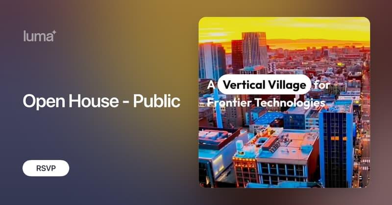
Toggle light / dark theme

Emotional responses to sensory experience are central to the human condition in health and disease. We hypothesized that principles governing the emergence of emotion from sensation might be discoverable through their conservation across the mammalian lineage. We therefore designed a cross-species neural activity screen, applicable to humans and mice, combining precise affective behavioral measurements, clinical medication administration, and brain-wide intracranial electrophysiology. This screen revealed conserved biphasic dynamics in which emotionally salient sensory signals are swiftly broadcast throughout the brain and followed by a characteristic persistent activity pattern. Medication-based interventions that selectively blocked persistent dynamics while preserving fast broadcast selectively inhibited emotional responses in humans and mice.

Erwin Schrödinger’s famous thought experiment has always been deeply misunderstood. In this article I’d like to explain how, if understood properly, it might shed new light on the mechanism by which consciousness evolved.

Schrödinger’s cat and schrödinger’s hat

The purpose of Schrödinger’s thought experiment was to highlight serious problems in the (then very new) “Copenhagen Interpretation” of Quantum Mechanics (CI). The CI was a bit of a botch-job, because the founders of QM had no idea how to “interpret” the strange new physics they had discovered. The CI says quantum systems remain in a superposition (a “smeared out” state where everything than can happen is somehow happening in parallel) until measured, but does not define what counts as a “measurement”, or why. Schrödinger always rejected this idea, and his thought experiment was intended to demonstrate why. He proposes a sealed box (so no “measurements” can take place), in which has been placed a cat, and a quantum source with a 50% probability of releasing poison. According to the CI, so long as the system inside the box remains “unmeasured”, the poison has both been released and not-released and therefore that cat is both dead and alive.

Narcissism has become the armchair diagnosis of the decade. Social media is awash with people flinging the label around. Everyone’s ex seems to be a narcissist, some of our parents are under suspicion, and that office villain? They definitely tick the box, too.

The accuracy of these rampant diagnoses warrants scepticism. But the reality is narcissists do exist. At its extreme, narcissism is a rare mental health diagnosis, known as narcissistic personality disorder. But narcissism also describes a cluster of personality traits, which we all display to varying degrees.

For those of us who have been in close quarters with someone high in narcissistic traits, we rarely walk away unscathed. And we may be left with lingering questions. For example, what made them this way?

In a diseased condition, most of the time, target proteins attain toxicity following their transition from a α-helix to a β-sheet form [18]. Although numerous functional native proteins possess β-sheet conformations within them, the transition from an α-helix to a β-sheet is characteristic of amyloid deposits [19], and often associated with the change of a physiological function to a pathological one. Such abnormal conformational transition exposes hydrophobic amino acid residues and promotes protein aggregation [18, 20]. The toxic proteins often interact with other native proteins and may catalyze their transition into a toxic sate, and hence they are called infective conformations [18]. The newly formed toxic proteins can repeat this cycle to intiate a self-sustaining loop; thereby amplifying the toxicity to generate a catastrophic effect, beyond homeostatic reparative mechanisms, to eventually impair cellular function or induce cellular demise [21].

Proteins function properly when their constituent amino acids fold correctly [22]. On the other hand, misfolded proteins assemble into insoluble aggregates with other proteins and can be toxic for the cells [18, 20]. Ataxin-1 is highly prone to misfolding due to inherited gene defects that cause neurodegenerative diseases (NDDs), which is mainly due the repetition of glutamine within its amino acid chain; the toxicity of this protein being directly proportional to the number of glutamines [23]. There are 21 proteins that mainly interact with ataxin-1 and influence its folding or misfolding, 12 of which increase the toxicity of ataxin-1 for nerve cells, while 9 of the identified proteins reduce its toxicity [23]. Ataxin-1 resembles a double twisted spiral or helix and has a special structure, termed a “coiled coil domain”, that promotes aggregation. Proteins which possess “coiled coil domain” and interact with ataxin-1 have been reported to enhance promotion of ataxin-1 aggregation and toxic effects [24].

The gradual accumulation of misfolded proteins in the absence of their appropriate clearance can cause amyloid disease, the most prevalent one being AD. Parkinson’s disease and Huntington’s disease have similar amyloid origins [25]. These diseases can be sporadic or familial and their incidence increases dramatically with age. The mechanistic explanation for this correlation is that as we age (and are subjected to increasing numbers of mutations and/or oxidative stress causing changes to protein structure, etc.), the delicate balance of the synthesis, folding, and degradation of proteins is disturbed, ensuing in the production, accumulation and aggregation of misfolded proteins [26].

People spend about a third of their lives asleep. Yet, surprisingly little is known about how our brains control falling asleep and waking up. Now, researchers led by Prof. Henrik Bringmann at the Biotechnology Center (BIOTEC) of TUD Dresden University of Technology discovered another piece of this puzzle. The team showed that a single brain signal acts like a biological switch—both triggering sleep and ending it.

Their findings, published in the journal Current Biology, were made possible by studying a tiny roundworm, C. elegans, a powerful model organism in biology.

“It is really important to be able to fall asleep, but just as important to wake up too,” says Prof. Bringmann, research group leader at BIOTEC who led the study.

A new study published in Nature reveals how olfactory sensory neurons (OSNs) achieve extraordinary precision in selecting which genes to express.

The mechanism is surprising in that it involves solid-like molecular condensates that last for days, helping to solve a long-standing puzzle in genome organization.

The research, led by Prof. Stavros Lomvardas from Columbia University, addresses one of biology’s most intriguing questions: How do in the nose manage to express only one (OR) gene out of approximately 1,000 available options?