Menu

Blog

Archive for the ‘neuroscience’ category: Page 260

Sep 8, 2023

Effects of electromagnetic fields exposure on the antioxidant defense system

Posted by in categories: biotech/medical, food, neuroscience

If you want to read a long and complex article on EMF and oxidative stress, here’s one. And when I did a Google search on my Pixel 6 that has generative results, the result said it affected the blood brain barrier but I think it combined several websites as a source and summarized them. I’ll post a screenshot.


Free radicals are reactive molecules produced during the conversion of foods into energy through oxygen. The formation of free radicals is an oxidation reaction that occurs on an oxygen basis. [27]. Since oxygen is essential for survival, the formation of free radicals cannot be avoided. However, factors including ionizing and non-ionizing radiation alter the transcription and translation of genes such as JUN, HSP 70 and MYC, via the epidermal growth factor receptor EGFR-ras, leading to the generation of ROS [28,29] and resulting in the overproduction of ROS in tissues [30].

The Fenton reaction is a catalytic process that converts hydrogen peroxide, a product of mitochondrial oxidative respiration, into a highly toxic hydroxyl free radical. Some studies have suggested that EMF is another mechanism through the Fenton reaction, suggesting that it promotes free radical activity in cells [31,32]. Although some researchers have reported that ROS perform beneficial function, a high degree of ROS production may cause cellular damage, resulting in a range of diseases. These radicals react with various biomolecules, including DNA ( Fig. 1 ). Namely, the energy of free radicals is not enough, and for this reason they behave like robbers who seize energy from other cells and rob a person to satisfy themselves [33]. Many studies have suggested that EMF may trigger the formation of reactive oxygen species in exposed cells in vitro [34,35,36,37] and in vivo [7, 31,38]. The initial stage of the ROS production in the presence of RF is controlled by the NADPH oxidase enzyme located in the plasma membrane. Consequently, ROS activate matrix metalloproteases, thereby initiating intracellular signaling cascades to warn the nucleus of the presence of external stimulation. These changes in transcription and protein expression are observed after RF exposure [39]. Kazemi et al. investigated the effect of exposure to 900-MHz on the induction of oxidative stress and the level of intracellular ROS in human mononuclear cells. Excessive elevation in ROS levels is an important cause of oxidative damage in lipids and proteins and nucleic acids. It therefore causes changes in enzyme activity and gene expression, eventually leading to various diseases, including sleep disorder, arthrosclerosis, loss of appetite, diabetes, dizziness, rheumatoid arthritis, cardiovascular disease, nausea and stroke [40,41,42]. In addition, degradation of the pro-oxidant-antioxidant balance due to an uncontrolled increase in ROS may also result in lipid peroxidation. Lipid peroxidation is the process in which cell membranes are rapidly destroyed due to the oxidation of components of phospholipids containing unsaturated fatty acids. By continuing this reaction, lipid peroxides (-C0, H) accumulate in the membrane, and transform polyunsaturated fatty acids into biologically active substances [43]. Consequently, lipid peroxidation leads to significant damage in the cells, such as disturbances in membrane transport, structural changes, cell membrane fluidity, damage to protein receptors in membrane structures, and changes in the activity of cell membrane enzymes [44]. Hoyto et al. demonstrated significant induction of lipid peroxidation after exposure to EMF in the mouse SH-SY5Y cell and L929 fibroblast cells [45]. Epidemiological studies have also suggested that oxidative damage to lipids in blood vessel walls may be a significant contributor to the development of atherosclerosis [46,47,48].

Sep 7, 2023

Brain functions best in a balanced state, new study claims

Posted by in categories: biotech/medical, computing, neuroscience

A new study explores the field of biological brain modeling, determining the brain’s structure is similar to a computer.

The brain has been designated as one of the most complex organs of the human body, comprised of features including intelligence, an interpreter of the senses, an initiator of body movement, and a controller of behavior, according to the National Institute of Health.

Now, a recent study discovered that a living model of this three-pound organ sheds light on the mechanisms of how humans understand and experience the world.

Sep 7, 2023

Is the Blood-Brain Barrier a Key to Behavior?

Posted by in categories: biotech/medical, neuroscience

Summary: The blood-brain barrier (BBB) in carpenter ants isn’t just a protective boundary, but actively shapes ant behavior.

The BBB produces an enzyme called Juvenile hormone esterase (Jhe) that degrades the Juvenile Hormone (JH3), which promotes foraging behavior. The presence and degradation of JH3 by the BBB helps determine whether an ant becomes a forager or soldier.

Interestingly, similar mechanisms might influence mouse behavior, hinting at broader implications beyond ants.

Sep 7, 2023

“Reality” is constructed by your brain. Here’s what that means, and why it matters

Posted by in categories: computing, cosmology, neuroscience, space travel

Year 2020 The ecology of the human brain is so complex that it even seems like it’s own not only story within itself but also could be like a self perpetuating universe of all sorts. Even neurons resemble the universe. What I believe is that the human brain is actually like an infinite spaceship that has infinite potential not only as a computational source but as sentience that is actual sentient in itself not just a story but kinda the god in the machine like a black box of limitless potential not only a computer but much more possibly a universe that guides us and shapes us. Even when we see the ecology of the mind we see so many stories and realities able to create its own multiverse… More.


What the science of visual illusions can teach us about our polarized world.

Sep 7, 2023

Researchers identify the link between memory and appetite in the human brain to explain obesity

Posted by in categories: biotech/medical, food, neuroscience

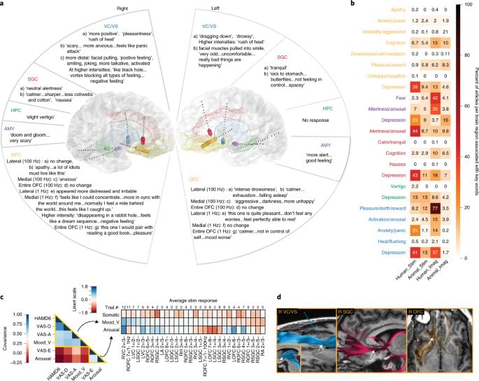
Disrupted connections between memory and appetite regulating brain circuits are directly proportional to body mass index (BMI), notably in patients who suffer from disordered or overeating that can lead to obesity, such as binge eating disorder (BED), according to new research from the Perelman School of Medicine at the University of Pennsylvania. Published today in Nature, the research notes that individuals who are obese have impaired connections between the dorsolateral hippocampus (dlHPC) and the lateral hypothalamus (LH), which may impact their ability to control or regulate emotional responses when anticipating rewarding meals or treats.

“These findings underscore that some individual’s brains can be fundamentally different in regions that increase the risk for obesity,” senior author, Casey Halpern, MD, an associate professor of Neurosurgery and Chief of Stereotactic and Functional Neurosurgery at Penn Medicine and the Corporal Michael J. Crescenz Veterans Affairs Medical Center. “Conditions like disordered eating and obesity are a lot more complicated than simply managing self-control and eating healthier. What these individuals need is not more willpower, but the therapeutic equivalent of an electrician that can make right these connections inside their brain.”

The dlHPC is located in the region of the brain that processes memory, and the LH is in the region of the brain that is responsible for keeping the body in a stable state, called homeostasis. Previous research has found an association with loss of function in the human hippocampus in individuals with obesity and related disordered eating, like BED. However, outside of imaging techniques such as magnetic resonance imaging (MRI), the role of the hippocampus has been difficult to study in humans with obesity and related eating disorders.

Sep 7, 2023

InnovationRx: New Insight Into How The Brain Works

Posted by in categories: chemistry, neuroscience

To understand why this is a big deal, for a long time its been understood that (to vastly oversimplify things) the brain is primarily composed to two kinds of cells: glial cells, which are basically the brain’s infrastructure; and neurons, which communicate with each other with chemicals called neurotransmitters at special sites called synapses.


InnovationRx is your weekly digest of healthcare news. To get it in your inbox, subscribe here.

Add another layer of complexity to our understanding of the brain. Researchers at University of Lausanne have discovered that a heretofore unknown class of cell is also involved in the complicated internal communications of the brain. The research was published Wednesday in Nature.

Continue reading “InnovationRx: New Insight Into How The Brain Works” »

Sep 7, 2023

Traumatic brain injuries linked to cognitive decline later in life

Posted by in category: neuroscience

A study of twins who served in World War II showed that traumatic brain injuries are associated with faster rates of cognitive decline as we age.

Sep 6, 2023

The discovery of a new kind of cell shakes up neuroscience

Posted by in category: neuroscience

A research team from University of Lausanne (UNIL) and the Wyss Center, has discovered a new type of cell essential for brain function. Hybrid in composition and function, in between the two types of brain cells known so far—the neurons and the glial cells—these cells of a new order are present in several brain regions in mice and humans.

The study published in the journal Nature shows that these cells promote the ability to memorize, the control of movements, and contrast the insurgence of epileptic seizures.

Neuroscience is in great upheaval. The two major families of cells that make up the brain, neurons and glial cells, secretly hid a hybrid cell, halfway between these two categories. For as long as neuroscience has existed, it has been recognized that the brain works primarily thanks to the neurons and their ability to rapidly elaborate and transmit information through their networks.

Sep 6, 2023

Breakthrough brain cell discovery shocks neuroscientists

Posted by in categories: health, neuroscience

Scientists have discovered a new type of brain cell that promises to shake up the field of neuroscience.

The discovery brings an end to a decades-old controversy and may pave the way for new targeted treatments for a range of health conditions.

Sep 6, 2023

Tattoo technique transfers gold nanopatterns onto live cells

Posted by in categories: biotech/medical, chemistry, cyborgs, nanotechnology, neuroscience, transhumanism

For now, cyborgs exist only in fiction, but the concept is becoming more plausible as science progresses. And now, researchers are reporting in ACS’ Nano Letters that they have developed a proof-of-concept technique to “tattoo” living cells and tissues with flexible arrays of gold nanodots and nanowires. With further refinement, this method could eventually be used to integrate smart devices with living tissue for biomedical applications, such as bionics and biosensing.

Advances in electronics have enabled manufacturers to make integrated circuits and sensors with nanoscale resolution. More recently, laser printing and other techniques have made it possible to assemble flexible devices that can mold to curved surfaces. But these processes often use harsh chemicals, high temperatures or pressure extremes that are incompatible with living cells. Other methods are too slow or have poor spatial resolution. To avoid these drawbacks, David Gracias, Luo Gu and colleagues wanted to develop a nontoxic, high-resolution, lithographic method to attach nanomaterials to living tissue and cells.

The team used nanoimprint lithography to print a pattern of nanoscale gold lines or dots on a polymer-coated silicon wafer. The polymer was then dissolved to free the gold nanoarray so it could be transferred to a thin piece of glass. Next, the gold was functionalized with cysteamine and covered with a hydrogel layer, which, when peeled away, removed the array from the glass. The patterned side of this flexible array/hydrogel layer was coated with gelatin and attached to individual live fibroblast cells. In the final step, the hydrogel was degraded to expose the gold pattern on the surface of the cells. The researchers used similar techniques to apply gold nanoarrays to sheets of fibroblasts or to rat brains. Experiments showed that the arrays were biocompatible and could guide cell orientation and migration.

Page 260 of 1,039First257258259260261262263264Last