Toggle light / dark theme

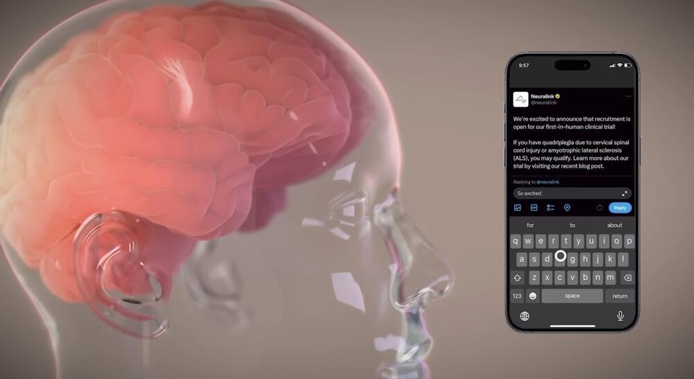
Neuralink, the Elon Musk-founded company developing implantable chips that can read brain waves, has raised an additional $43 million in venture capital, according to a filing with the SEC.

The filing published this week shows the company increased its previous tranche, led by Peter Thiel’s Founders Fund, from $280 million to $323 million in early August. Thirty-two investors participated, according to the filing.

Neuralink hasn’t disclosed its valuation recently. But in June, Reuters reported that the company was valued at about $5 billion after privately-executed stock trades.

5th BigBrain Workshop 2021
22 September 2021 — Applications.
Chair: Kathleen Rockland.

The Unique Cytoarchitecture and Wiring of The Default Mode Network.
Casey Paquola.

Background. Complex behaviours benefit from parallel distributed processing in multiple brain networks. The roles of certain networks are well-defined, while others remain elusive. Arguably, none are so elusive as the default mode network (DMN); a distributed set of brain regions that decrease in activity during many externally oriented tasks. Revealing the cytoarchitectural composition and connectional layout of the DMN is crucial to defining its role in complex behaviours.

Method. We examined the cytoarchitectural composition of the DMN using an established cortical type atlas (García-Cabezas et al., 2020; Von Economo and Koskinas, 1925) and by applying non-linear dimensionality reduction to BigBrain-derived staining intensity profiles (Paquola et al., 2019). Next, we used magnetic resonance imaging (MRI) to explicate structural wiring and effective connectivity of the whole brain. In both modalities, we examined the influence of cytoarchitecture on extrinsic connectivity of the DMN. Finally, we evaluated the uniqueness of the DMN relative to other large-scale functional brain networks.

Results. We discovered profound diversity of DMN cytoarchitecture. Each circumscribed subregion of the DMN contains a broad range of cytoarchitectural types, however, the spatial pattern within each subregion differs. The patterns vary in smoothness from a gradient in the parahippocampus to interdigitation in the superior frontal gyrus. We found that cytoarchitectural differentiation in the DMN aligns with its structural wiring and extrinsic information flow. The structural heterogeneity of the DMN engenders a network-level balance in communication with external and internal sources, which is distinctive, relative to other functional networks.
Conclusion. These findings suggest a novel wiring diagram of structural and functional connectivity of the DMN that is compatible with its putative role in balancing internal and external information. Furthermore, our work demonstrates the import of neuroanatomical evidence in specifying theories of functional networks.

All information about the 5th BigBrain Workshop 2021, including detailed authors information: https://go.fzj.de/BigBrainWorkshop2021

An amazing graph theoretic analysis of the C. elegans neuropeptide connectome!


Neuromodulation by peptides is essential for brain function. By comprehensively mapping neuropeptide signaling in the nematode C. elegans, Ripoll-Sánchez et al. define a dense wireless network whose organization differs in important ways from wired brain circuits. This network is a prototype for understanding neuropeptide signaling networks in larger brains.

To try everything Brilliant has to offer—free—for a full 30 days, visit http://brilliant.org/ArtemKirsanov/
The first 200 of you will get 20% off Brilliant’s annual premium subscription.

My name is Artem, I’m a computational neuroscience student and researcher. In this video we discuss engrams – fundamental units of memory in the brain. We explore what engrams are, how memory is allocated, where it is stored, and how different memories become linked with each other.

Patreon: https://www.patreon.com/artemkirsanov.
Twitter: https://twitter.com/ArtemKRSV

OUTLINE:
00:00 — Introduction.
00:39 — Historical background.
01:44 — Fear conditioning paradigm.
03:38 — Immediate-early genes as memory markers.
08:13 — Engrams are necessary and sufficient for recall.
10:16 — Excitabiliy and memory allocation.
16:19 — Brain-wide engrams.
18:12 — Linking memories together.
24:20 — Summary.
25:33 — Brilliant.
27:09 — Outro.

REFERENCES (in no particular order):
1. Robins, S. The 21st century engram. WIRES Cognitive Science e1653 (2023) doi:10.1002/wcs.1653.
2. Roy, D. S. et al. Brain-wide mapping reveals that engrams for a single memory are distributed across multiple brain regions. Nat Commun 13, 1799 (2022).
3. Josselyn, S. A. & Tonegawa, S. Memory engrams: Recalling the past and imagining the future. Science 367, eaaw4325 (2020).
4. Chen, L. et al. The role of intrinsic excitability in the evolution of memory: Significance in memory allocation, consolidation, and updating. Neurobiology of Learning and Memory 173, 107266 (2020).
5. Rao-Ruiz, P., Yu, J., Yu, J. J., Kushner, S. A. & Josselyn, S. A. Neuronal competition: microcircuit mechanisms define the sparsity of the engram. Current Opinion in Neurobiology 54163–170 (2019).
6. Josselyn, S. A. & Frankland, P. W. Memory Allocation: Mechanisms and Function. Annu. Rev. Neurosci. 41389–413 (2018).
7. Choi, J.-H. et al. Interregional synaptic maps among engram cells underlie memory formation. Science 360430–435 (2018).
8. Abdou, K. et al. Synapse-specific representation of the identity of overlapping memory engrams. Science 360, 1227–1231 (2018).
9. Yokose, J. et al. Overlapping memory trace indispensable for linking, but not recalling, individual memories. Science 355398–403 (2017).
10. Rashid, A. J. et al. Competition between engrams influences fear memory formation and recall. Science 353383–387 (2016).
11. Poo, M. et al. What is memory? The present state of the engram. BMC Biol 14, 40 (2016).
12. Park, S. et al. Neuronal Allocation to a Hippocampal Engram. Neuropsychopharmacol 41, 2987–2993 (2016).
13. Morrison, D. J. et al. Parvalbumin interneurons constrain the size of the lateral amygdala engram. Neurobiology of Learning and Memory 135, 91–99 (2016).
14. Minatohara, K., Akiyoshi, M. & Okuno, H. Role of Immediate-Early Genes in Synaptic Plasticity and Neuronal Ensembles Underlying the Memory Trace. Front. Mol. Neurosci. 8, (2016).
15. Josselyn, S. A., Köhler, S. & Frankland, P. W. Finding the engram. Nat Rev Neurosci 16521–534 (2015).
16. Yiu, A. P. et al. Neurons Are Recruited to a Memory Trace Based on Relative Neuronal Excitability Immediately before Training. Neuron 83722–735 (2014).
17. Redondo, R. L. et al. Bidirectional switch of the valence associated with a hippocampal contextual memory engram. Nature 513426–430 (2014).
18. Ramirez, S. et al. Creating a False Memory in the Hippocampus. Science 341387–391 (2013).
19. Liu, X. et al. Optogenetic stimulation of a hippocampal engram activates fear memory recall. Nature 484381–385 (2012).
20. Silva, A. J., Zhou, Y., Rogerson, T., Shobe, J. & Balaji, J. Molecular and Cellular Approaches to Memory Allocation in Neural Circuits. Science 326391–395 (2009).

CREDITS:

Cleveland Clinic researchers analyzed genes and brain tissue of patients with Alzheimer’s and found that differences in brain immunometabolism – the interactions between the immune system and the ways cells create energy – may contribute to women’s increased risk for the disease and its severity.

The findings, published in Alzheimer’s and Dementia, offer important insight into developing sex-specific treatment and prevention options for Alzheimer’s disease, the sixth-leading cause of death in the United States.

“Our immune systems depend on communication between different cell types in our bodies, which are fueled by energy created from unique metabolic processes,” said Justin Lathia, Ph.D., vice chair of the Department of Cardiovascular and Metabolic Sciences and co-author on the paper. “As sex influences both the immune system and metabolic process, our study aimed to identify how all of these individual factors influence one another to contribute to Alzheimer’s disease.”

Contrary to popular belief, the brain does not have the capability to rewire itself to compensate for loss of sight, amputations, or stroke-related damage, according to scientists from the University of Cambridge and Johns Hopkins University.

In a recent paper published in eLife, Professors Tamar Makin (Cambridge) and John Krakauer (Johns Hopkins) argue that the notion that the brain, in response to injury or deficit, can reorganize itself and repurpose particular regions for new functions, is fundamentally flawed – despite being commonly cited in scientific textbooks. Instead, they argue that what is occurring is merely the brain being trained to utilize already existing, but latent, abilities.

Link : https://trib.al/DwqJQ8E


Researchers in Texas have developed a method to keep a brain alive and functioning for several hours without being connected to the body — a truly weird scientific experiment that recalls the head in jars bit in the iconic cartoon “Futurama.”

A team led by the University of Texas Southwestern Medical Center in Dallas took two pigs and severed connections between their heads and bodies, instead hooking the brains up to a device they call the extracorporeal pulsatile circulatory control (EPCC), which they detailed in a paper published in the journal Scientific Reports. The machine keeps blood pumping through the brain, mimicking the natural flow when it’s connected to the rest of the body.

The intent behind this nightmarish procedure was to study the brain independently from other bodily functions that may influence it, but the system may also lead to better-designed cardiopulmonary bypass, a process in which machines take over your heart and lung function during surgery.

A recent study has raised questions about the impact of chronic caffeine consumption on our brain’s ability to adapt and learn. In a new study published in Frontiers in Psychiatry, scientists found that long-term caffeine users may exhibit decreased brain plasticity, a critical factor in the processes of learning and memory, when subjected to a brain stimulation protocol.

Caffeine is a common stimulant found in coffee, tea, soda, and other beverages. It’s known to help with alertness and concentration, but its effects on the brain’s ability to change and adapt over time, a process called plasticity, have been less clear.

Previous studies have shown mixed results when it comes to caffeine’s influence on brain plasticity. Some studies hinted that caffeine might hinder the brain’s ability to learn and adapt, while others suggested potential benefits. The researchers conducted this study to investigate the effects of caffeine on human brain plasticity, specifically focusing on its impact on long-term potentiation (LTP) and its potential interaction with a neuromodulation technique called repetitive transcranial magnetic stimulation (rTMS).