Toggle light / dark theme

Year 2023 face_with_colon_three


Bioengineers and tissue engineers intend to reconstruct skin equivalents with physiologically relevant cellular and matrix architectures for basic research and industrial applications. Skin pathophysiology depends on skin-nerve crosstalk and researchers must therefore develop reliable models of skin in the lab to assess selective communications between epidermal keratinocytes and sensory neurons.

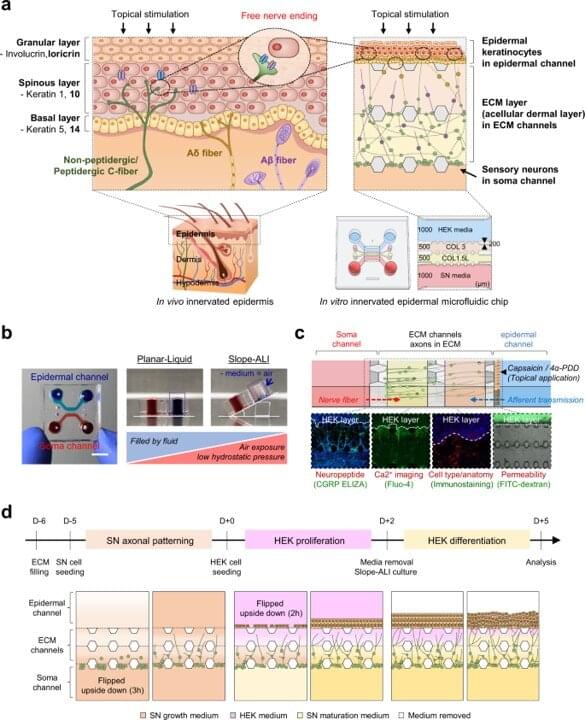
In a new report now published in Nature Communications, Jinchul Ahn and a research team in , bio-convergence engineering, and therapeutics and biotechnology in South Korea presented a three-dimensional, innervated epidermal keratinocyte layer on a to create a sensory neuron-epidermal keratinocyte co-culture model. The maintained well-organized basal-suprabasal stratification and enhanced barrier function for physiologically relevant anatomical representation to show the feasibility of imaging in the lab, alongside functional analyses to improve the existing co-culture models. The platform is well-suited for biomedical and pharmaceutical research.

Skin: The largest sensory organ of the human body

Skin is composed of a complex network of sensory nerve fibers to form a highly sensitive organ with mechanoreceptors, thermoreceptors and nociceptors. These neuronal subtypes reside in the dorsal root ganglia and are densely and distinctly innervated into the cutaneous layers. Sensory nerve fibers in the skin also express and release nerve mediators including neuropeptides to signal the skin. The biological significance of nerves to sensations and other biological skin functions have formed physical and pathological correlations with several skin diseases, making these instruments apt in vivo models to emulate skin-nerve interactions.

Medication delivered by a novel gel cured 100% of mice with an aggressive brain cancer, a striking result that offers new hope for patients diagnosed with glioblastoma, one of the deadliest and most common brain tumors in humans.


Cui’s team combined an anticancer drug and an antibody in a solution that self-assembles into a gel to fill the tiny grooves left after a brain tumor is surgically removed. The gel can reach areas that surgery might miss and current drugs struggle to reach to kill lingering cancer cells and suppress tumor growth. The results are published in Proceedings of the National Academy of Sciences.

The gel also seems to trigger an immune response that a mouse’s body struggles to activate on its own when fighting glioblastoma. When the researchers rechallenged surviving mice with a new glioblastoma tumor, their immune systems alone beat the cancer without additional medication. The gel appears to not only fend off cancer but help rewire the immune system to discourage recurrence with immunological memory, researchers said.

Still, surgery is essential for this approach, the researchers said. Applying the gel directly in the brain without surgical removal of the tumor resulted in a 50% survival rate.

In a recent study published in Molecular Psychiatry, researchers explored the effects of a small humanin-like peptide 2 (SHLP2) variant on mitochondrial function.

Mitochondria are implicated in Parkinson’s disease (PD) pathogenesis. Mitochondrial-derived peptides (MDPs) are microproteins encoded from small open reading frames (sORFs) in the mitochondrial DNA (mtDNA). SHLP2 is an MDP with an essential role in multiple cellular processes, and it improves mitochondrial metabolism by increasing biogenesis and respiration and reducing oxidation.

Recent studies link mitochondrial single nucleotide polymorphisms (mtSNPs) within coding regions of MDPs to age-related deficits. For instance, m.2706 A G, an mtSNP in humanin, predicts reduced circulating levels of humanin and worse cognitive decline. Moreover, another mtSNP, m.2158 T C, is associated with reduced PD risk, albeit the underlying mechanisms are unknown.

How does the developing brain process surprising sounds and what changes as we grow up?


For children, the world is full of surprises. Adults, on the other hand, are much more difficult to surprise. And there are complex processes behind this apparently straightforward state of affairs. Researchers at the University of Basel have been using mice to decode how reactions to the unexpected develop in the growing brain.

Babies love playing peekaboo, continuing to react even on the tenth sudden appearance of their partner in the game. Recognizing the unexpected is an important cognitive ability. After all, new can also mean dangerous.

The exact way in which surprises are processed in the as we grow, however: unusual stimuli are much more quickly categorized as “important” or “uninteresting,” and are significantly less surprising the second and third time they appear. This increased efficiency makes perfect sense: new stimuli may gain our attention, but do not cause an unnecessarily strong reaction that costs us energy. While this may appear trivial at first, so far there has been very little research into this fact in the context of development.

The neural processes behind memory encoding in the brain have been revealed in this new research.


However, beneath this seemingly effortless experience, there exist intricate neural processess.

Dartmouth College researchers have identified the complicated neurological systems that regulate how the human brain stores memories.

The findings revealed a “neural coding mechanism” in the brain that allows information to be transferred between perception and memory regions.

Year 2021 face_with_colon_three


In our new paper, we’ve investigated how quantum particles could move in a complex structure like the brain, but in a lab setting. If our findings can one day be compared with activity measured in the brain, we may come one step closer to validating or dismissing Penrose and Hameroff’s controversial theory.

Brains and Fractals

Our brains are composed of cells called neurons, and their combined activity is believed to generate consciousness. Each neuron contains microtubules, which transport substances to different parts of the cell. The Penrose-Hameroff theory of quantum consciousness argues that microtubules are structured in a fractal pattern which would enable quantum processes to occur.

Our memories are rich in detail: we can vividly recall the color of our home, the layout of our kitchen, or the front of our favorite café. How the brain encodes this information has long puzzled neuroscientists.

In a new Dartmouth-led study, researchers identified a neural coding mechanism that allows the transfer of information back and forth between perceptual regions to memory areas of the . The results are published in Nature Neuroscience.

Prior to this work, the classic understanding of brain organization was that perceptual regions of the brain represent the world “as it is,” with the brain’s visual cortex representing the external world based on how light falls on the retina, “retinotopically.” In contrast, it was thought that the brain’s memory areas represent information in an abstract format, stripped of details about its physical nature. However, according to the co-authors, this explanation fails to take into account that as information is encoded or recalled, these regions may in fact, share a common code in the brain.