For researchers wanting to understand the inner workings of magnetic materials, transmission electron microscopy is an indispensable tool. Because the wavelength of an electron is much shorter than the wavelength of visible light, a beam of electrons transmitted through a thin slice of a material can create an image in which the inner structure of the material is magnified up to 50 million times, many orders of magnitude more than with an optical microscope.
Since transmission electron microscopy, or TEM, was first developed in the 1930s, scientists and engineers have come up with many variations on the technique. Each of these variations suit different applications and come with their own advantages and disadvantages.
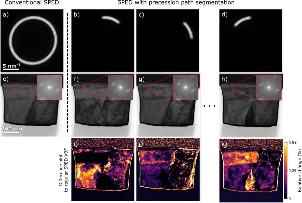
Now, researchers at NTNU have come up with a remarkably simple method to solve a long-standing problem in an almost 30-year-old TEM-based technique known as scanning precession electron diffraction, or SPED.


Comments are closed.